. 2013 Aug 25;376(1-2):156-72.
doi: 10.1016/j.mce.2013.06.016. Epub 2013 Jun 20.

Exposure to chemical cocktails before or after conception--- the effect of timing on ovarian development

Affiliations

Exposure to chemical cocktails before or after conception--- the effect of timing on ovarian development

Michelle Bellingham et al. Mol Cell Endocrinol. .

Abstract

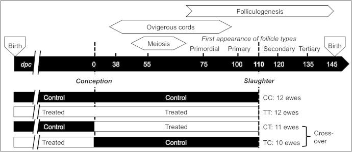
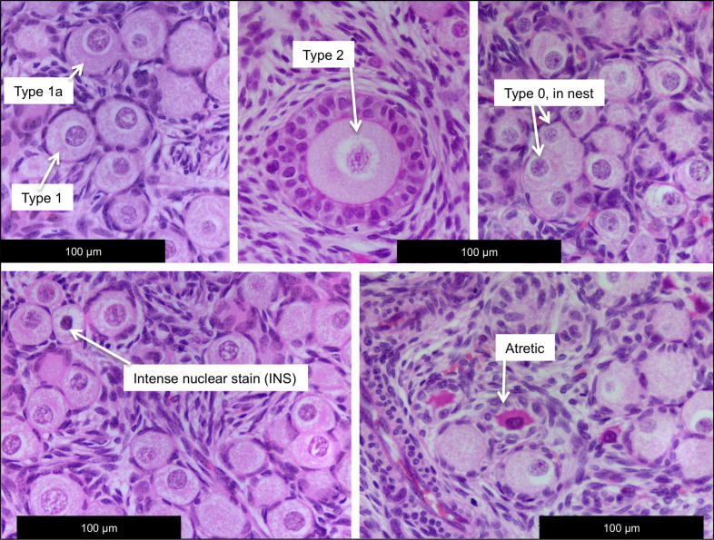
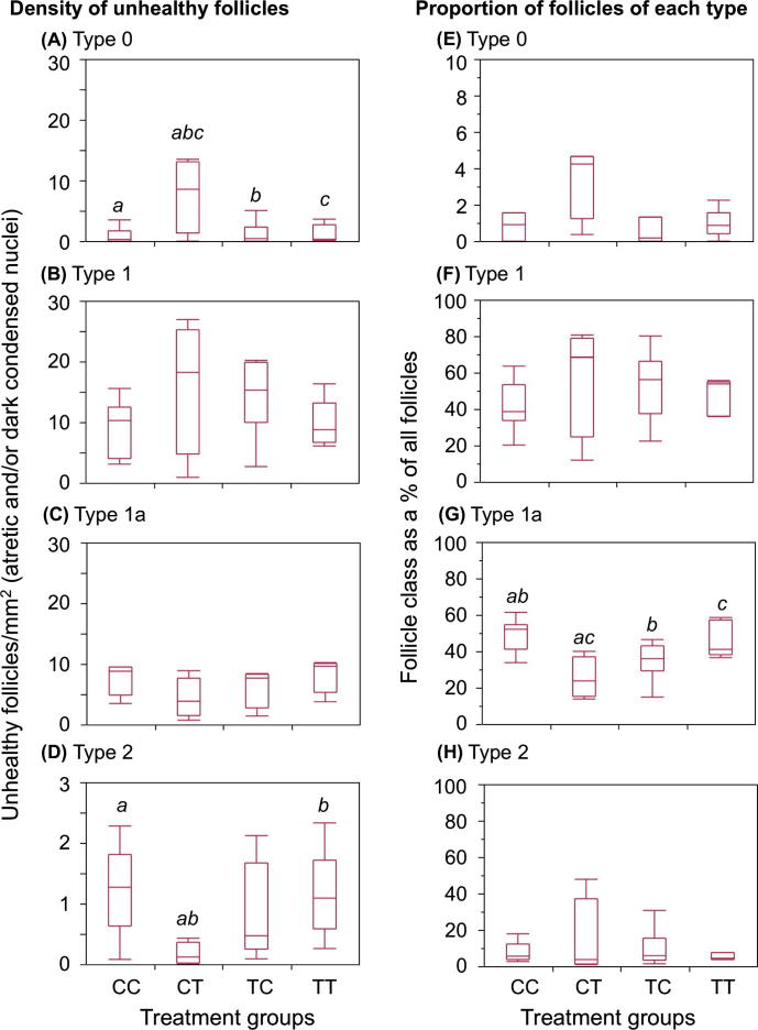
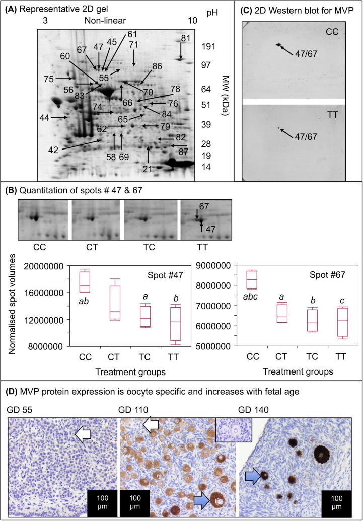
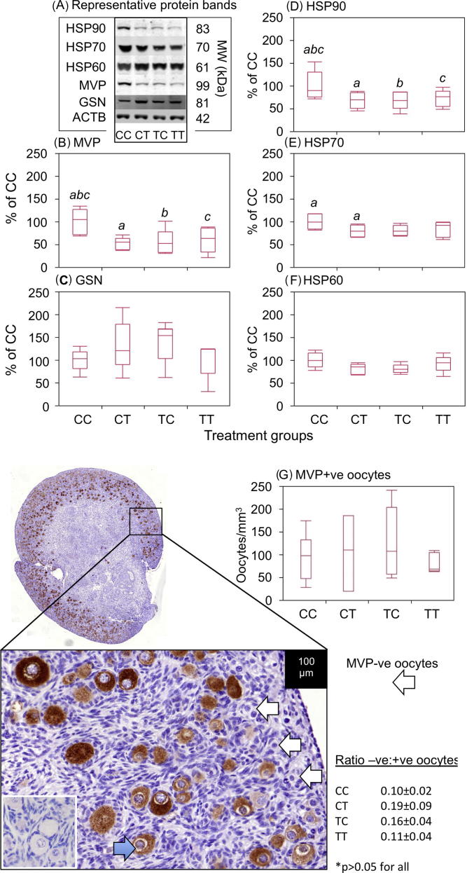
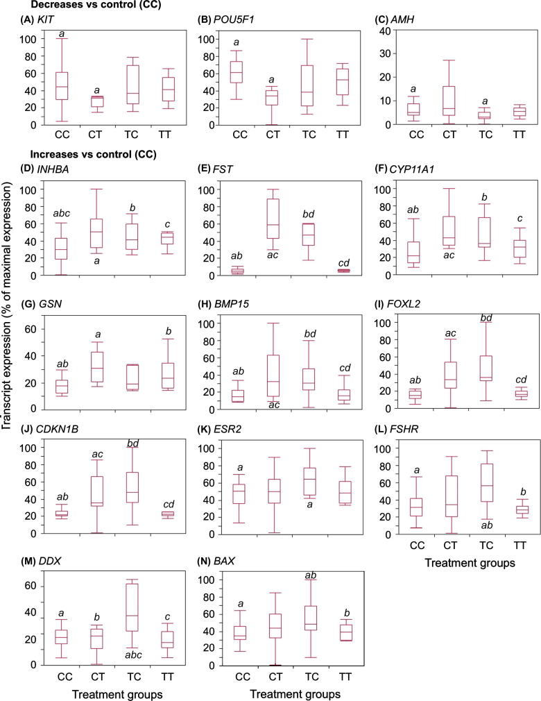
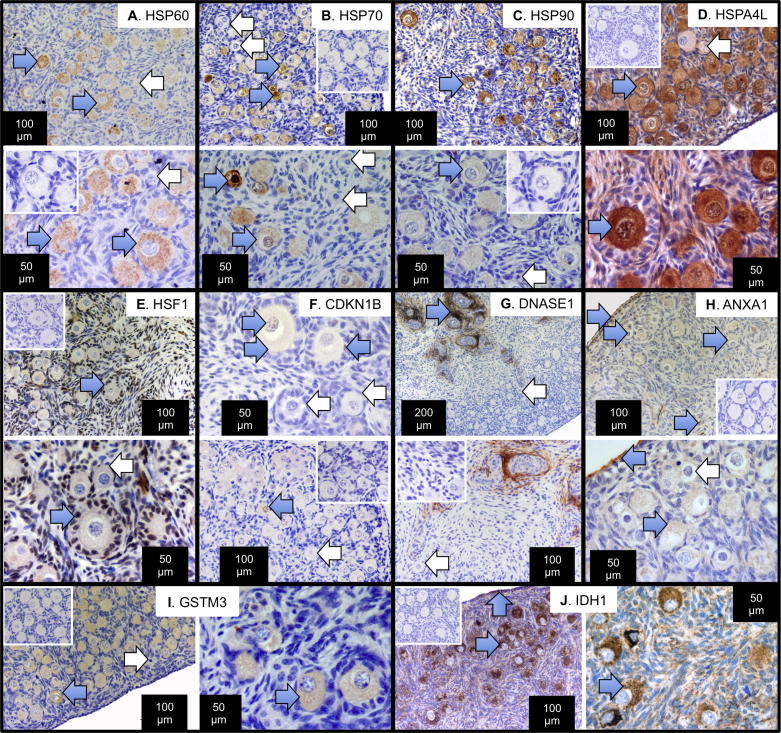
Exposure of female fetuses to environmental chemicals (ECs) during pregnancy results in a disturbed ovarian adult phenotype. We investigated the influence of pre- and/or post-conception exposure to low-level mixtures of ECs on the structure and function of the fetal ovine ovary. We examined ovarian morphology, expression of oocyte and granulosa cell-specific genes and proteome. Female fetuses were collected at day 110 of gestation, from dams exposed continuously until, and after mating, by grazing in pastures treated with sewage sludge as a fertiliser (TT) or in control fields treated with inorganic fertiliser (CC). In addition, in a cross-over design, fetal ovaries were collected from dams maintained on sludge pastures up to the time of mating but then transferred to control pastures (TC) and, reciprocally, those transferred from control to treated pastures at mating (CT). On examination, the proportion of type 1a follicles (activating primordial follicles) was significantly lower in animals from the CT groups compared with CC and TT groups (P<0.05). Of the 23 ovarian gene transcripts studied, 14 were altered in the ovaries of exposed fetuses (CT, TC, and TT) relative to controls, with the largest number of changes observed in cross-exposure pattern groups (CT or TC). Continuous EC exposure (TT) produced fewer transcript alterations and only two genes (INHBA and GSN) presented differential profiles between CC and TT. Fetal ovarian proteome analysis (2-DE gels) showed, across all exposure groups, 86 differentially expressed protein spots compared to controls. Animals in the CT group exhibited the highest number (53) while TC and TT presented the same number of affected protein spots (42). Fetal ovarian proteins with altered expression included MVP (major vault protein) and several members of the heat-shock family (HSPA4L, HSP90AA1 and HSF1). The present findings indicate that continuous maternal EC exposure before and during gestation, are less deleterious for fetal ovarian development than a change in maternal EC exposure between pre and post-conception. The pathways by which the ovary responds to this chemical stress were common in TT, CT, TC exposed foetuses. In addition to the period of pregnancy, the pre-conception period appears also as crucial for conditioning long-term effects of EC exposure on ovarian development and primordial follicle reserve and hence future fertility.

Keywords: Anti-ACTB; DEHP; Development; ECs; EDCs; Environmental chemicals; FSH; In utero exposure; LH; Mixtures; Ovary; WB; Western blot; anti-β actin; diethylhexylphthalate; endocrine disrupting chemicals; environmental chemicals; follicle stimulating hormone; luteinising hormone.


Figures

Fig. 1
Fig. 1
Diagrammatic summary of study design. Adults ewes were maintained on either control (inorganic fertiliser) or exposed (sewage sludge fertiliser) pastures until mating The CC and TT ewes continued on the same pastures, but the cross-over ewes, TC, CT, were moved to the opposite exposure pastures. The CC ewes were never exposed to sewage sludge while the TT ewes were always exposed to sewage sludge. The TC ewes were exposed to sewage sludge only before mating while the CT ewes were only exposed to sewage sludge after mating.
Fig. 2
Fig. 2
Representative oocyte and follicle classification used to quantify the morphological effects of sewage sludge exposures on the fetal sheep ovary. The figure shows typical examples of oogonial nests, types 0, 1, 1a and 2 follicles (healthy) and also intense nuclear staining and follicular atresia.
Fig. 3
Fig. 3
Exposure to sewage sludge from conception onwards alters fetal ovarian follicle characteristics. The densities of unhealthy follicles (A–D) classed as those with condensed, intensely stained nuclei and/or atresia are shown combined, while the relative proportions of all follicles of each class, relative to total follicle density (E–H) are shown in box and whisker plots. The horizontal line in the boxes show the median values, with the limits of the boxes showing the 25% and 75% quantiles and the whiskers showing the 10% and 90% quantiles. Common superscripts between groups, for each follicle type, denote statistically significant differences at P < 0.05.
Fig. 4
Fig. 4
Representative proteomic data. (A) Representative 2-DE gel of CC group ovarian proteins is shown, with the spots identified in Table 3 denoted by arrows and numbers. (B) Quantitation of spots 47 and 67, both identified as MVP. Representative zoom-boxes for each treatment group are shown, as well as box and whisker plots of normalised spot volumes. The horizontal lines in the boxes show the median values, with the limits of the boxes showing the 25% and 75% quantiles and the whiskers showing the 10% and 90% quantiles. Common superscripts between groups, for each protein spot, denote statistically significant differences at P < 0.05. (C) Representative, not-quantitative 2-DE Western blots for MVP showing the localisation of a single spot at the locus of spot #47. D. Representative immunohistochemistry showing that MVP expression increases dramatically between day 55 and 140 of gestation in the fetal sheep ovary and is oocyte-specific. The inset box shows IgG-ve staining control.
Fig. 5
Fig. 5
Sewage sludge exposure affects MVP, HSP90 and HSP70 proteins in fetal ovaries, quantified by Western blot. (A) Representative bands for each treatment group for each Western blot, including β-actin load control. These bands are all from the same 4 ovaries and all available ovaries were used for these Western blots. (B–F) Quantitation of 5 ovarian proteins shown as box and whisker plots. The band volume for each protein was normalised against β-actin for the same lane (i.e. same ovary) and then expressed relative to the mean normalised band volume of the CC treatment group. The horizontal line in the boxes show the median values, with the limits of the boxes showing the 25% and 75% quantiles and the whiskers showing the 10% and 90% quantiles. Common superscripts between groups, for each protein, denote statistically significant differences at P < 0.05. Where there are no superscripts p values are >0.05. (G) Quantification of MVP immunopositive and immunonegative oocytes shows that there were no significant exposure effects on the density of MVP immunopositive oocytes or the ratio between immunonegative and immunopositive oocytes, although the latter tended to be higher in the CT and TC groups.
Fig. 6
Fig. 6
Sewage sludge exposure affects expression of developmental and reproductive mRNA transcripts. (A–C) show transcripts with significant reduction compared with controls while (D–N) show transcript with significant increase in expression relative to controls (CC). Transcript expression was determined by qPCR, normalised relative to the house-keeping gene HPRT1, expressed relative to maximal expression levels for each transcript separately and shown as box and whisker plots. The horizontal line in the boxes show the median values, with the limits of the boxes showing the 25% and 75% quantiles and the whiskers showing the 10% and 90% quantiles. For each transcript separately, common superscripts between groups denote statistically significant differences at P < 0.05.
Fig. 7
Fig. 7
Immunolocalisation of proteins identified as affected by sewage sludge exposure in the fetal sheep ovary. Heat-shock proteins, HSP60 (A), HSP70 (B), HSP90 (C) and HSPA4L (D) were all predominantly oocyte-specific, with strong cytoplasmic staining in most oocytes (blue arrows) but not all oocytes (white arrows). The fifth heat-shock protein, HSF1 (E) was localised in granulosa cells, pre-granulosa cells and many (but not all) somatic cells around the follicles. At higher power, HSF1 expression was detected in the nuclei of granulosa cells (blue arrows), but not in all pre-granulosa cells around primordial or forming primordial follicles (white arrow). CDKN1B (F) was localised in the cytoplasm and/or nuclei of some but not all oocytes and in the nuclei of some granulosa cells (blue arrows). Atretic follicles were CDKN1B-negative, but so were some healthy oocytes and follicles (white arrows). DNASE1 (G) showed intense staining in somatic cells around blood vessels and mesenchymal remnants (blue arrow) although all oocytes were negative (white arrow). ANXA1 (H) exhibited quite wide-spread staining, especially in the ovarian surface epithelium, the cytoplasm of many oocytes and scattered somatic cells (blue arrows). GSTM3 (I) was also principally localised to oocyte cytoplasm (blue arrow) although some oocytes, particularly those showing signs of atresia or dark condensed nuclei were negative (white arrow). IDH1 (J) was localised mainly in oocyte cytoplasm (blue arrow) but also in some somatic cells, including some surface epithelium cells. Positive staining is brown (DAB), counterstained by haematoxylin (blue). The bars denote scale for each image separately. Blue arrows highlight immuno-positive cells and white arrows immuno-negative cells. Two magnifications, taken from different ovaries, are shown for each antigen, separated by a white line. Each antigen is bounded by a black box to simplify interpretation. In all cases IgG-negative slides incubated with non-immune serum of the appropriate species were characterised by an absence of brown stain (one inset panel for each antigen).

References

    1. Abbondanza C., Rossi V., Roscigno A., Gallo L., Belsito A., Piluso G., Medici N., Nigro V., Molinari A.M., Moncharmont B., Puca G.A. Interaction of vault particles with estrogen receptor in the MCF-7 breast cancer cell. J. Cell Biol. 1998;141:1301–1310. - PMC - PubMed
    1. Adhikari D., Liu K. Molecular mechanisms underlying the activation of mammalian primordial follicles. Endocr. Rev. 2009;30:438–464. - PubMed
    1. Alcorn J., McNamara P.J. Ontogeny of hepatic and renal systemic clearance pathways in infants: Part II. Clin. Pharmacokinet. 2002;41:1077–1094. - PubMed
    1. Alcorn J., McNamara P.J. Pharmacokinetics in the newborn. Adv. Drug Deliv. Rev. 2003;55:667–686. - PubMed
    1. Bellingham M., Fowler P.A., Amezaga M.R., Rhind S.M., Cotinot C., Mandon-Pepin B., Sharpe R.M., Evans N.P. Exposure to a complex cocktail of environmental endocrine-disrupting compounds disturbs the kisspeptin/GPR54 system in ovine hypothalamus and pituitary gland. Environ. Health Perspect. 2009;117:1556–1562. - PMC - PubMed

Publication types

MeSH terms