

Aspergilloma mimicking a lung cancer
- PMID: 23792483
- PMCID: PMC3710896
- DOI: 10.1016/j.ijscr.2013.02.028
Aspergilloma mimicking a lung cancer
Abstract
Introduction: Pulmonary aspergillosis occurs in the parenchymal cavities or ectatic airways. It rarely affects healthy people with an intact immune response. There have been few reports describing an aspergilloma mimicking a lung cancer.
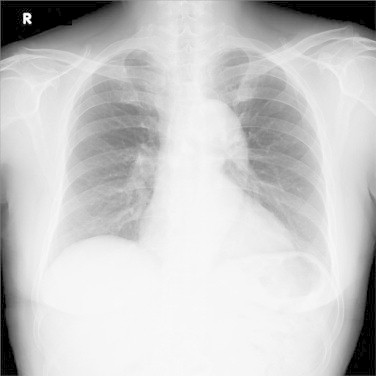
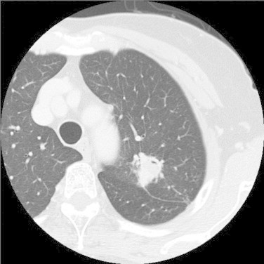
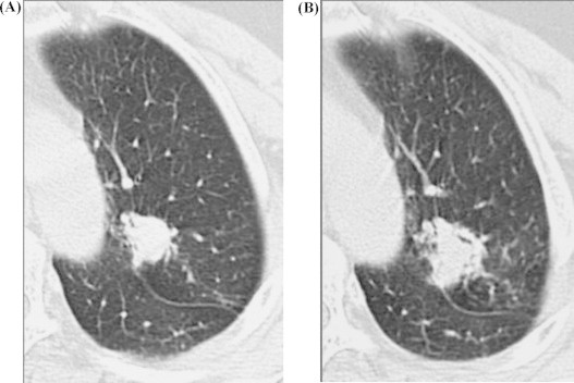
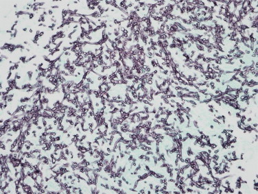
Presentation of case: We experienced the case of an asymptomatic healthy 71-year-old female who was admitted with an abnormal lung shadow. Chest CT revealed an irregularly shaped solid lung nodule in the left upper lobe, which increased in size during the follow-up at a regional hospital. The pathology of the bronchial biopsy was negative for malignant cells, and the cultures were negative. Because a lung cancer was strongly suspected, video-assisted thoracic surgery was performed. Aspergillus was detected by a pathological study of the excised specimen, with no evidence of lung cancer.
Discussion: It is difficult to make an accurate diagnosis of aspergilloma by imaging findings in healthy people with an intact immune response, and therefore a surgical resection allows both the pathological diagnosis and treatment to be performed concurrently.
Conclusion: An aspergilloma presenting a mass shadow on imaging may mimic a lung cancer in healthy people with intact immune response.
Keywords: Aspergilloma; Imaging findings; Lung cancer.
Copyright © 2013. Published by Elsevier Ltd.
Figures
References
-
- Gefter W.B. The spectrum of pulmonary aspergillosis. Journal of Thoracic Imaging. 1992;7:56–74. - PubMed
-
- Aquino S.L., Lee S.T., Warnock M.L., Gamsu G. Pulmonary aspergillosis: imaging findings with pathologic correlation. American Journal of Roentgenology. 1994;163:811–815. - PubMed
-
- Park Y., Kim T.S., Yi C.A., Cho E.Y., Kim H., Choi Y.S. Pulmonary cavitary mass containing a mural nodule: differential diagnosis between intracavitary aspergilloma and cavitating lung cancer on contrast-enhanced computed tomography. Clinical Radiology. 2007;62:227–232. - PubMed
-
- Yoon S.H., Park C.M., Goo J.M., Lee H.J. Pulmonary aspergillosis in immunocompetent patients without air-meniscus sign and underlying lung disease: CT findings and histopathologic features. Acta Radiologica. 2011;52:756–761. - PubMed
-
- Lowe V.J., Fletcher J.W., Gobal L., Lawson M., Kirchner P., Valk P. Prospective investigation of positron emission tomography in lung nodules. Journal of Clinical Oncology. 1998;16:1075–1084. - PubMed
LinkOut - more resources
Full Text Sources
Other Literature Sources
Research Materials
