. 2013 Jul;31(7):653-8.
doi: 10.1038/nbt.2614. Epub 2013 Jun 23.

Efficiency of siRNA delivery by lipid nanoparticles is limited by endocytic recycling

Affiliations

Efficiency of siRNA delivery by lipid nanoparticles is limited by endocytic recycling

Gaurav Sahay et al. Nat Biotechnol. 2013 Jul.

Abstract

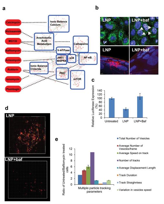
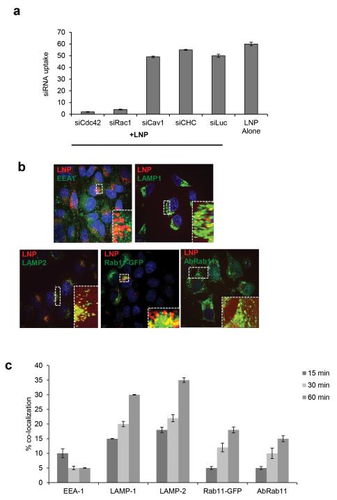
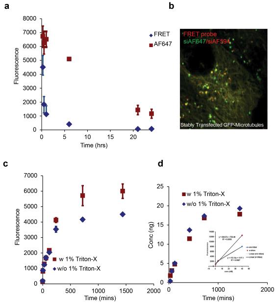
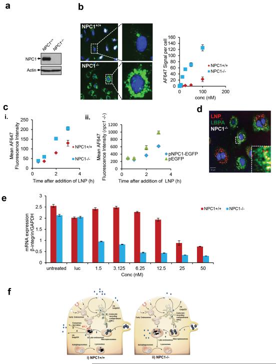
Despite efforts to understand the interactions between nanoparticles and cells, the cellular processes that determine the efficiency of intracellular drug delivery remain unclear. Here we examine cellular uptake of short interfering RNA (siRNA) delivered in lipid nanoparticles (LNPs) using cellular trafficking probes in combination with automated high-throughput confocal microscopy. We also employed defined perturbations of cellular pathways paired with systems biology approaches to uncover protein-protein and protein-small molecule interactions. We show that multiple cell signaling effectors are required for initial cellular entry of LNPs through macropinocytosis, including proton pumps, mTOR and cathepsins. siRNA delivery is substantially reduced as ≅70% of the internalized siRNA undergoes exocytosis through egress of LNPs from late endosomes/lysosomes. Niemann-Pick type C1 (NPC1) is shown to be an important regulator of the major recycling pathways of LNP-delivered siRNAs. NPC1-deficient cells show enhanced cellular retention of LNPs inside late endosomes and lysosomes, and increased gene silencing of the target gene. Our data suggest that siRNA delivery efficiency might be improved by designing delivery vehicles that can escape the recycling pathways.


Figures

Figure 1
Figure 1. Systems survey for endocytosis of lipid nanoparticles (LNP)
LNPs containing siAF647 were incubated with HeLa-GFP cells (50nM) for 3 hrs in the presence or absence of small molecules and imaged using an automated high throughput confocal microscope a. Small molecules that inhibit over 80% of internalization (with no more than 10%-15% loss of cell viability) and their network interactions are presented in the form of a systems diagram b. Localization of siAF647LNP in presence or absence of bafilomycin is represented as an individual image (arrows indicate peripheral localization) and a 3D z-stack. c. Dual HeLa cells were exposed to LNP (siLuc, 10nM) in the presence or absence of bafilomycin (1μM). Luciferase to Renilla levels was measured for silencing activity. Experiments were performed in triplicate; errors are plotted as standard error means (S.E.M.). (d-e). Time lapse Total internal reflection (TIRF) microscopy of cells exposed to siAF647-LNPs (50nM, 3hrs) in the presence or absence of bafilomycin was subjected to multiple particle tracking (MPT). A snapshot of vesicular tracks indicating the movement of LNPs in presence or absence of bafilomycin presented in (d). The ratio between the MPT parameters for untreated/bafilomycin treated cells was calculated to provide a quantitative measure of differences in LNP trafficking with or without bafilomycin (e). Errors of ratios are plotted through propagation of errors from division.
Figure 2
Figure 2. Cellular trafficking of LNPs
a. Quantitative image analysis of siAF647-LNP cellular uptake (3 hrs) in HeLa cells silenced with siRNA against key endocytic regulators (Cdc42, Rac-1, Clathrin heavy chain (CHC), caveolin-1 (Cav-1). siRNA against luciferase serve as a negative control. (b-c) Image based quantitative analysis of siAF647-LNPs co-localization (3 hrs pulse, 15, 30, 60 min. chase) with markers of endocytosis, anti-EEA-1 (early endosomes), anti-LAMP-1 (late endosome/lysosomes), anti-LAMP-2 (late endosomes/lysosomes), Rab11-GFP and anti-Rab11 (both mark endocytic recycling compartment). High-resolution z-stack confocal images representative of 60 min chase are presented in different panels (b). Quantification of LNP-siRNA cellular uptake after indicated times (a, c).
Figure 3
Figure 3. Quantitative analysis for disassembly and recycling of LNPs
a. HeLa cells were exposed to LNP with the FRET pair (AF647/AF594 siRNA), washed and replenished with media. Changes in FRET (excitation-561 nm, emission-641 nm) intensity were monitored at different time points using flow cytometery to quantify LNP disassembly. The emission from a single fluorophore (excitation-633 nm, emission-641 nm) was used to measure intracellular siRNA at these time points. b. Cellular uptake of FRET probe-LNP (siAF647/siAF594, 3hrs) was imaged in stably transfected GFP-tubulin cells 1 hour post incubation c. siAF647-LNP was pulsed for 3 hrs, washed and incubated with fresh media to remove non internalized particles. Media was removed at multiple time points and analyzed using a fluorescent reader to determine amount recycled. 1% Triton-X was later added (red curve) to the media and fluorescence was re-measured. d. Amount of siRNA was obtained from fluorescence values from (c) that were extrapolated using the standard curve (inset).
Figure 4
Figure 4. Enhanced cellular retention and efficacy of siAF647-LNPs in NPC1 deficient MEFs
a. Immunoblot analysis with anti-NPC1 antibody was used to validate wild type and NPC1 deficient MEFs. b. Automated confocal microscopy on NPC1+/+ or NPC1/ MEFs exposed to different concentrations of labeled LNPs and imaged 24 hrs post incubation. Inset from a representative image (100 nM) shows siRNA accumulation in individual cells. c. Flow cytometery analysis on LNP-siRNA uptake in (i) NPC1+/+ or NPC1/ cells or in (ii) NPC1/ cells transfected with pEGFP or pNPC1-GFP. The mean fluorescent intensity represents LNP uptake. d. NPC1/ cells treated with LNP-AF647-siRNA (red) (3 hrs pulse, 30 min chase) and immuno-stained with anti-LBPA antibody (green) e. LNPs containing siRNA against β integrin were added to wild type or NPC1 deficient MEFs as in (a), mRNA levels were quantitated at 24 hrs post incubation using branched DNA analysis. The experiment was done in triplicate and the errors are reported as S.E.M. f. A schematic representation of LNP trafficking (i) in NPC1+/+ and (ii) NPC1/ cells. Intact cationic LNPs enter through macropinocytosis (1); a small fraction of LNPs transport from macropinosomes to the endocytic recycling compartment (ERC) (2) while the majority is directed to late endosomes (3). Late endosome sort LNPs to lysosomes for degradation or utilize multiple recycling pathways to traffic them to the extracellular milieu. These mechanisms include recycling through transport to the ER-Golgi route (4) or direct fusion of late endosomes containing multivesicular bodies, with the plasma membrane (Exosomes secretion) (5). In NPC1 deficient cells the late endosome recycling mechanisms are impaired causing LNP-siRNA to accumulate in enlarged late endosomes leading to persistent escape of siRNA that improves gene silencing. (Intact nanoparticle-formula image, siRNA complex-formula image)

Comment in

References

    1. Elbashir SM, et al. Duplexes of 21-nucleotide RNAs mediate RNA interference in cultured mammalian cells. Nature. 2001;411:494–8. - PubMed
    1. Whitehead KA, Langer R, Anderson DG. Knocking down barriers: advances in siRNA delivery. Nat Rev Drug Discov. 2009;8:129–138. - PMC - PubMed
    1. Davis ME, et al. Evidence of RNAi in humans from systemically administered siRNA via targeted nanoparticles. Nature. 2010;464:1067–1070. - PMC - PubMed
    1. Sheridan C. Proof of concept for next-generation nanoparticle drugs in humans. Nature Biotechnology. 2012;30:471–473. - PubMed
    1. Sahay G, Alakhova DY, Kabanov AV. Endocytosis of nanomedicines. J Control Release. 2010;145:182–195. - PMC - PubMed

Publication types

MeSH terms