. 2013 Aug 2;288(31):22555-64.
doi: 10.1074/jbc.M113.482356. Epub 2013 Jun 21.

Amino-terminal domain tetramer organization and structural effects of zinc binding in the N-methyl-D-aspartate (NMDA) receptor

Affiliations

Amino-terminal domain tetramer organization and structural effects of zinc binding in the N-methyl-D-aspartate (NMDA) receptor

Rita E Sirrieh et al. J Biol Chem. .

Abstract

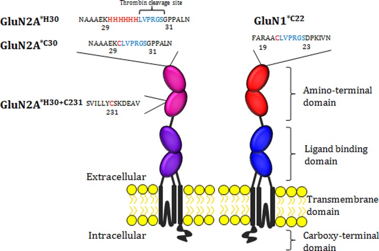
N-Methyl-D-aspartate (NMDA) receptors mediate excitatory neurotransmission in the mammalian central nervous system. An important feature of these receptors is their capacity for allosteric regulation by small molecules, such as zinc, which bind to their amino-terminal domain (ATD). Zinc inhibition through high affinity binding to the ATD has been examined through functional studies; however, there is no direct measurement of associated conformational changes. We used luminescence resonance energy transfer to show that the ATDs undergo a cleft closure-like conformational change upon binding zinc, but no changes are observed in intersubunit distances. Furthermore, we find that the ATDs are more closely packed than the related AMPA receptors. These results suggest that the stability of the upper lobe contacts between ATDs allow for the efficient propagation of the cleft closure conformational change toward the ligand-binding domain and transmembrane segments, ultimately inhibiting the channel.

Keywords: Allosteric Regulation; Amino-terminal Domain; Fluorescence Resonance Energy Transfer (FRET); Glutamate Receptors Ionotropic (AMPA, NMDA); Ion Channels; LRET; Zinc.


Figures

FIGURE 1.
FIGURE 1.
Comparison of intersubunit distances of the AMPA and NMDA receptor ATDs. A, crystal structure of the ATDs from the full-length GluA2 homotetramer is shown (PDB ID code 3KG2 (7)). The B and D subunits are in green, and the A and C subunits are in blue. Stated distances are measured from the α-carbon of Asn-10, shown in red. B, top view of the GluN1/GluN2B isolated ATD structure is shown (PDB ID code 3QEL (6)). The GluN2B subunits (corresponding to the B and D subunits of GluA2) are colored green, and the GluN1 subunits (conforming to the A and C subunits) are colored blue. Distances are measured from the α-carbon of Pro-24 of GluN1 and Pro-32 of GluN2B, shown in red.
FIGURE 2.
FIGURE 2.
Schematic of an individual iGluR subunit showing label sites for LRET studies. The amino acid sequence and number for the label sites with the construct name are shown in addition to the labeled domains of the receptor. Residues that bind fluorophores are highlighted in red with thrombin cleavage sites shown in blue.
FIGURE 3.
FIGURE 3.
Zinc sensitivity of constructs used for LRET. A, representative whole cell recording from CHO cell expressing GluN1*/GluN2A*H30+C231 receptors in response to 100 μm glutamate alone or with 3, 30, or 300 nm free zinc. B, summary zinc inhibition curves for each construct recorded at 50 mV with 4–8 cells for each point. Note that each construct shows high affinity zinc block with an IC50 between 17 and 45 nm zinc, except for the zinc-insensitive GluN2A*H30+C231/DHM mutant (red circles). Error bars, S.E.
FIGURE 4.
FIGURE 4.
Lifetime measurements of NMDA receptor intersubunit distances in CHO cells. A, GluN2A-GluN2A LRET lifetimes obtained by co-expressing GluN1* and GluN2A*C30 and labeling with terbium and Alexa Fluor 555 as donor and acceptor fluorophores, respectively. B, GluN1-GluN2A LRET lifetimes labeled with terbium and Ni(NTA)2Cy3 as donor and acceptor fluorophores, respectively. C, GluN1-GluN1 LRET measurements labeled with terbium and Alexa Fluor 555 as donor and acceptor fluorophores, respectively. D, donor-only lifetimes for the GluN2A-GluN2A measurements. E, donor-only lifetimes for measurements between GluN1-GluN2A and GluN1-GluN1. In all panels, the black line represents lifetimes from receptors in extracellular buffer, green shows Tricine-buffered solutions, blue illustrates solutions with saturating zinc, and pink represents solutions with both saturating zinc and glutamate/glycine. All measurements can be fit with a single exponential lifetime decay curve.
FIGURE 5.
FIGURE 5.
Lifetime measurements of intersubunit distances in NMDA receptors expressed in X. laevis oocytes. A, GluN2A-GluN2A LRET lifetimes from co-expressed GluN1* and GluN2A*C30 labeled with terbium chelate and Alexa Fluor 555. B, GluN1-GluN2A LRET lifetimes measured from GluN1*C22 and GluN2A*H30 when labeled with terbium chelate and Ni(NTA)2Cy3. C, GluN1-GluN1 LRET lifetimes obtained from GluN1*C22 co-expressed with GluN2A*H30 and labeled with terbium chelate and Alexa Fluor 555. D, lifetimes for donor-labeled GluN2A*C30 used to calculate GluN2-GluN2 distances. E, donor-only lifetimes for GluN1*C22-labeled receptors to calculate GluN1-GluN2A and GluN1-GluN1 distances. In all panels, the black line represents lifetimes from receptors in extracellular buffer, green shows Tricine-buffered solutions, blue illustrates solutions with saturating zinc, and pink represents solutions with both saturating zinc and glutamate/glycine. All measurements can be fit with a single exponential lifetime decay curve.
FIGURE 6.
FIGURE 6.
Plot of energy transfer efficiency as a function of distance for terbium and Alexa Fluor 555. The red lines indicate the efficiency of energy transfer for the distance between GluN1 subunits in the GluN1-GluN2B ATD tetramer crystal structure (PDB ID code 3QEL (6)).
FIGURE 7.
FIGURE 7.
LRET lifetime measurements of the GluN2A ATD cleft. A, LRET lifetimes measured across the intrasubunit ATD cleft of GluN2A, obtained by co-expressing GluN1* and GluN2A*H30+C231 and labeling with terbium chelate and Ni(NTA)2Cy3. B, same lifetime measurement as in A performed on a zinc-insensitive GluN2A with mutations H44A and H128S. This measurement was obtained by co-expressing GluN1* and GluN2A*H30+C231/DHM and labeling with terbium chelate and Ni(NTA)2Cy3. C and D, donor-only lifetimes for the cleft measurements from constructs in A and B, respectively. In all panels, the black line represents lifetimes from receptors in extracellular buffer, green shows Tricine-buffered solutions, blue illustrates solutions with saturating zinc, and pink represents solutions with both saturating zinc and glutamate/glycine.

References

    1. Traynelis S. F., Wollmuth L. P., McBain C. J., Menniti F. S., Vance K. M., Ogden K. K., Hansen K. B., Yuan H., Myers S. J., Dingledine R. (2010) Glutamate receptor ion channels: structure, regulation, and function. Pharmacol. Rev. 62, 405–496 - PMC - PubMed
    1. Meddows E., Le Bourdelles B., Grimwood S., Wafford K., Sandhu S., Whiting P., McIlhinney R. A. (2001) Identification of molecular determinants that are important in the assembly of N-methyl-d-aspartate receptors. J. Biol. Chem. 276, 18795–18803 - PubMed
    1. Gielen M., Siegler Retchless B., Mony L., Johnson J. W., Paoletti P. (2009) Mechanism of differential control of NMDA receptor activity by NR2 subunits. Nature 459, 703–707 - PMC - PubMed
    1. Paoletti P., Perin-Dureau F., Fayyazuddin A., Le Goff A., Callebaut I., Neyton J. (2000) Molecular organization of a zinc binding N-terminal modulatory domain in an NMDA receptor subunit. Neuron 28, 911–925 - PubMed
    1. Masuko T., Kashiwagi K., Kuno T., Nguyen N. D., Pahk A. J., Fukuchi J., Igarashi K., Williams K. (1999) A regulatory domain (R1–R2) in the amino terminus of the N-methyl-d-aspartate receptor: effects of spermine, protons, and ifenprodil, and structural similarity to bacterial leucine/isoleucine/valine binding protein. Mol. Pharmacol. 55, 957–969 - PubMed

Publication types

LinkOut - more resources