. 2013 Dec:83:103-19.
doi: 10.1016/j.neuroimage.2013.06.042. Epub 2013 Jun 20.

Disrupted cortico-cerebellar connectivity in older adults

Affiliations

Disrupted cortico-cerebellar connectivity in older adults

Jessica A Bernard et al. Neuroimage. 2013 Dec.

Abstract

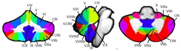
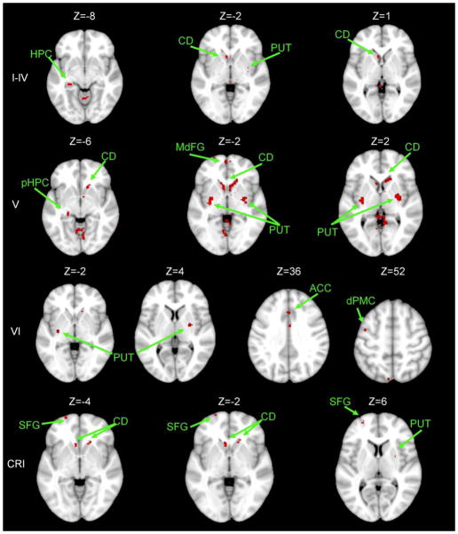
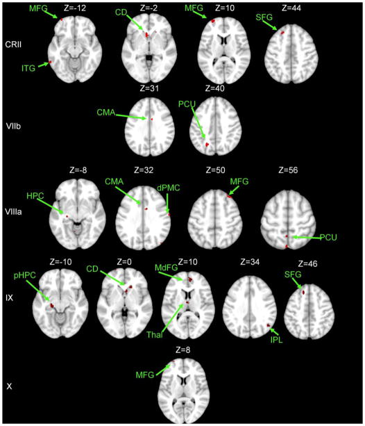
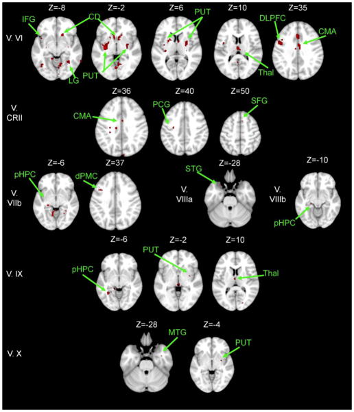
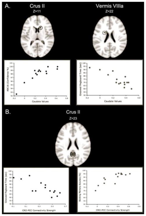
Healthy aging is marked by declines in a variety of cognitive and motor abilities. A better understanding of the aging brain may aid in elucidating the neural substrates of these behavioral effects. Investigations of resting state functional brain connectivity have provided insights into pathology, and to some degree, healthy aging. Given the role of the cerebellum in both motor and cognitive behaviors, as well as its known volumetric declines with age, investigating cerebellar networks may shed light on the neural bases of age-related functional declines. We mapped the resting state networks of the lobules of the right hemisphere and the vermis of the cerebellum in a group of healthy older adults and compared them to those of young adults. We report disrupted cortico-cerebellar resting state network connectivity in older adults. These results remain even when controlling for cerebellar volume, signal-to-noise ratio, and signal-to-fluctuation noise ratio. Specifically, there was consistent disruption of cerebellar connectivity with both the striatum and the medial temporal lobe. Associations between connectivity strength and both sensorimotor and cognitive task performances indicate that cerebellar engagement with the default mode network and striatal pathways is associated with better performance for older adults. These results extend our understanding of the resting state networks of the aging brain to include cortico-cerebellar networks, and indicate that age differences in network connectivity strength are important for behavior.

Keywords: Aging; Cerebellum; Medial temporal lobe; Sensorimotor performance; Striatum; Working memory; fcMRI.


Figures

Figure 1
Figure 1. Masks used as seed regions for connectivity analysis
The 17 lobular regions, as defined by the SUIT atlas (Diedrichsen, 2006; Diedrichsen et al., 2009) overlaid on a coronal (left), midsaggital (center), and axial (left) slices. Labels indicate the right hemisphere and vermal seeds used in this analysis. Adapted from Bernard and Seidler (In Press), Figure 1.
Figure 2
Figure 2. Areas exhibiting greater connectivity with cerebellar lobules I – Crus I in young adults versus older adults
Axial slices are presented, with the left hemisphere presented on the left. All results are thresholded using an uncorrected p<.001, and the clusters contain at least 10 voxels. CD: caudate; dPMC: dorsal pre-motor cortex; HPC: hippocampus; pHPC: parahippocampal gyrus; PUT: putamen; SFG: superior frontal gyrus.
Figure 3
Figure 3. Areas exhibiting greater connectivity with cerebellar lobules Crus II – X in young adults versus older adults
Axial slices are presented, with the left hemisphere presented on the left. All results are thresholded using an uncorrected p<.001, and the clusters contain at least 10 voxels.. CD: caudate; CMA: cingulate motor area; dPMC: dorsal pre-motor cortex; HPC: hippocampus; IPL: inferior parietal lobule; ITG: inferior temporal gyrus; MdFG: medial frontal gyrus; MFG: middle frontal gyrus; PCU: precuneus; pHPC: parahippocampal gyrus; SFG: superior frontal gyrus; Thal: thalamus.
Figure 4
Figure 4. Areas exhibiting greater connectivity with lobules of the cerebellar vermis in young adults versus older adults
Axial slices are presented, with the left hemisphere presented on the left. All results are thresholded using an uncorrected p<.001, and the clusters contain at least 10 voxels. CD: caudate; CMA: cingulate motor area; DLPFC: dorso-lateral prefrontal cortex; dPMC: dorsal pre-motor cortex; HPC: hippocampus; MTG: middle temporal gyrus; PCG: pre-central gyrus; pHPC: parahippocampal gyrus; IFG: inferior frontal gyrus; LG: lingual gyrus; PUT: putamen; SFG: superior frontal gyrus; STG: superior temporal gyrus; Thal: Thalamus.
Figure 5
Figure 5. Functional connectivity and behavior in older adults
A. Representative correlations between sensorimotor performance and connectivity strength between the cerebellum and caudate. Connectivity between Crus II and the caudate was positively correlated with balance confidence, while connectivity between Vermis lobule VIIIa was negatively correlated with the time taken to complete the Grooved pegboard. Stronger connectivity was associated with better task performance. B. Crus II connectivity with the posterior cingulate predicts sensorimotor and cognitive behavioral performance. Representative example correlations between Crus II and PCC connectivity strength and time to complete the grooved pegboard (left), and working memory accuracy (right). Greater connectivity strength was associated with better task performance. CRII: Crus II; PCC: Posterior-cingulate cortex.

References

    1. Allen G, Barnard H, McColl R, Hester AL, Fields JA, Weiner MF, Cullum CM. Reduced hippocampal functional connectivity in Alzheimer disease. Archives of Neurology. 2007;64:1482–1487. - PubMed
    1. Andrews-Hanna JR, Snyder AZ, Vincent JL, Lustig C, Head D, Raichle ME, Buckner RL. Disruption of large-scale brain systems in advanced aging. Neuron. 2007;56:924–935. - PMC - PubMed
    1. Anguera JA, Reuter-Lorenz PA, Willingham DT, Seidler RD. Contributions of spatial working memory to visuomotor learning. Journal of Cognitive Neuroscience. 2009;22:1917–1930. - PubMed
    1. Anguera JA, Reuter-Lorenz PA, Willingham DT, Seidler RD. Failure to engage spatial working memory contributes to age-related declines in visuomotor learning. Journal of Cognitive Neuroscience. 2011;22:1917–1930. - PubMed
    1. Avants BB, Epstein CL, Grossman M, Gee JC. Symmetric diffeomorphic image registration with cross-correlation: evaluating automated labeling of elderly and nerodegenerative brain. Medical Image Analysis. 2008;12:26–41. - PMC - PubMed

Publication types

MeSH terms