. 2013 Dec;36(6):1304-15.
doi: 10.1007/s10753-013-9669-0.

Changes in the expression of smooth muscle contractile proteins in TNBS- and DSS-induced colitis in mice

Affiliations

Changes in the expression of smooth muscle contractile proteins in TNBS- and DSS-induced colitis in mice

Reem Alkahtani et al. Inflammation. 2013 Dec.

Abstract

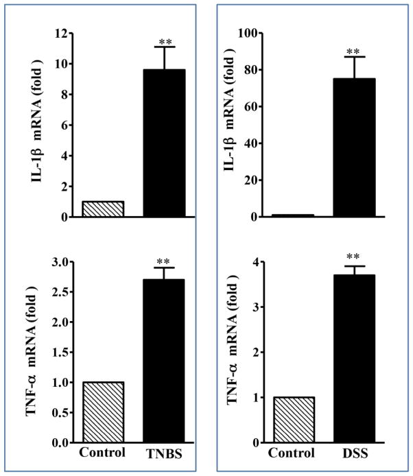
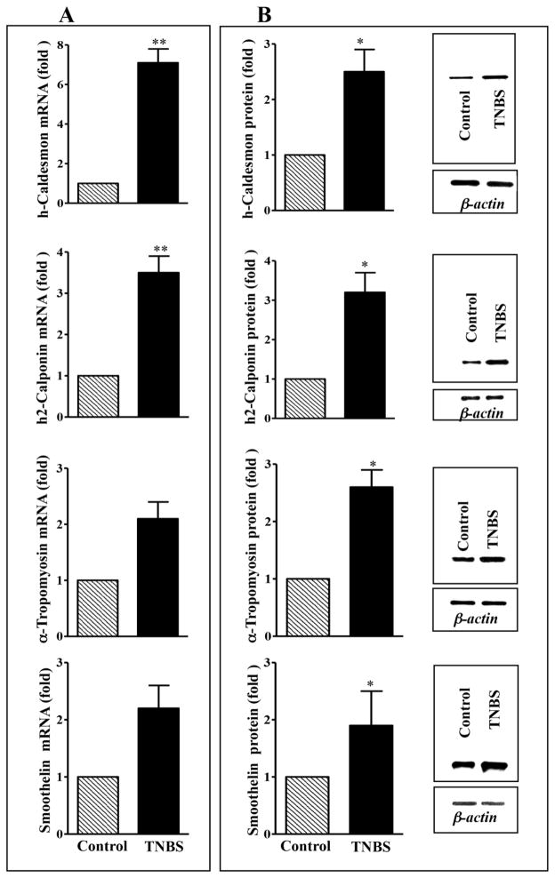
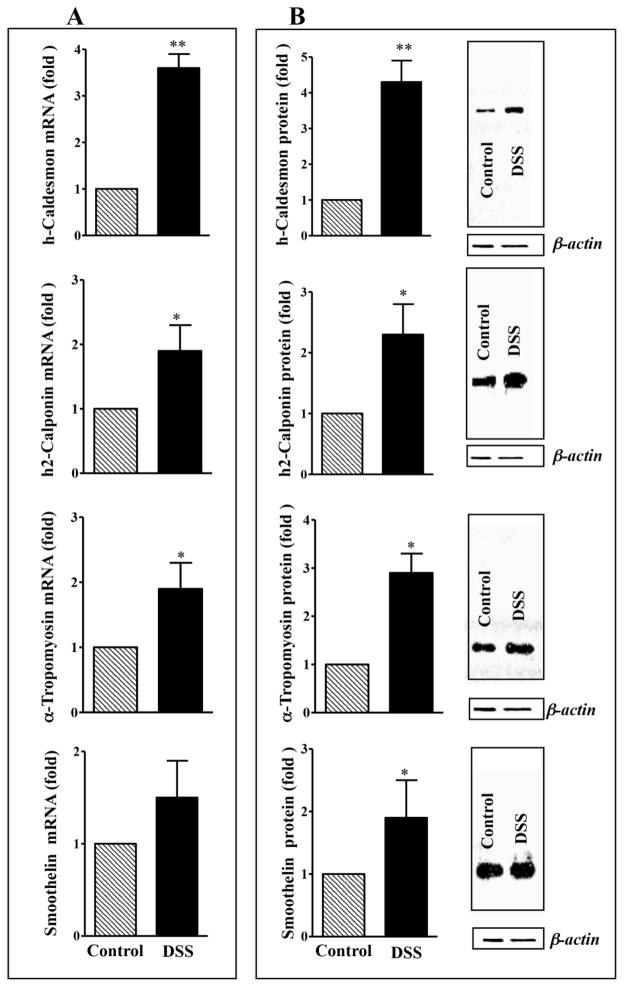
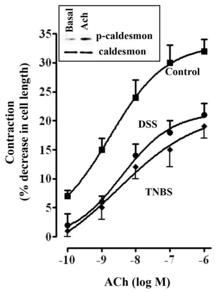
Thin filament-associated proteins such as calponin, caldesmon, tropomyosin, and smoothelin are thought to regulate acto-myosin interaction and thus, muscle contraction. However, the effect of inflammation on the expression of thin filament-associated proteins is not known. The aim of the present study is to determine the changes in the expression of calponin, caldesmon, tropomyosin, and smoothelin in colonic smooth muscle from trinitrobenzene sulphonic acid (TNBS)- and dextran sodium sulphate (DSS)-induced colitis in mice. Expression of h-caldesmon, h2-calponin, α-tropomyosin, and smoothelin-A was measured by qRT-PCR and Western blot. Contraction in response to acetylcholine in dispersed muscle cells was measured by scanning micrometry. mRNA and protein expression of α-actin, h2-calponin, h-caldesmon, smoothelin, and α-tropomyosin in colonic muscle strips from mice with TNBS- or DSS-induced colitis was significantly increased compared to control animals. Contraction in response to acetylcholine was significantly decreased in muscle cells isolated from inflamed regions of TNBS- or DSS-treated mice compared to control mice. Our results show that increase in the expression of thin filament-associated contractile proteins, which inhibit acto-myosin interaction, could contribute to decrease in smooth muscle contraction in inflammation.


Conflict of interest statement

Conflict of Interest Statement

The authors declare that there are no conflicts of interest.

Figures

Fig. 1
Fig. 1. Expression of IL-1β and TNF-α in the colonic muscle after TNBS or DSS treatment
Total RNA was isolated from smooth muscle strips of control and TNBS or DSS treated mice using RNAqueous prep kits (Ambion, Austin, TX) and was reverse transcribed using 2 μg of total RNA using qScript cDNA prep kits (Quanta, Gaithersburg, MD). The cDNA was amplified with specific primers for IL-1β or TNF α. The sequences of specific primers are listed in Table 1 and 2. Quantitative real-time polymerase chain reaction (qRT-PCR) was used to measure RNA levels of IL-1β or TNF α. For each cDNA sample, real-time PCR was conducted in a 20-μl reaction volume containing Quantitect SYBRgreen PCR Mastermix (Qiagen, Mississauga, ON). Real-time PCR reactions were performed in triplicate. Results are expressed as fold differences in IL-1β or TNF α gene expression in TNBS- or DSS-treated colon relative to that in vehicle-treated colon. Values represent the means ±SEM of 5 separate experiments. **p<0.001 versus control.
Fig. 2
Fig. 2. Expression of h-caldesmon, h2-calponin α-tropomyosin and smoothelin in the colon of control and TNBS-treated mice
(A) mRNA expression. Total RNA isolated from smooth muscle strips of control and inflamed colonic regions of TNBS-treated mice using RNAqueous prep kit and was reverse transcribed using 2 μg of total RNA using qScript cDNA prep kits. The cDNA was amplified with specific primers for h-caldesmon, h2-calponin, α-tropomyosin and smoothelin. The sequences of specific primers are listed in Table 1. For each cDNA sample, real-time PCR was conducted in a 20 μl reaction volume containing Taqman Gene expression PCR Mastermix. Real-time PCR reactions were performed in triplicate. Results are expressed as fold differences in h-caldesmon, h2-calponin, α-tropomyosin and smoothelin gene expression in TNBS-treated colon relative to that in control colon. Values represent the means ±SEM of 5 separate experiments. **p<0.001 versus control. (B) Protein expression. Colonic muscle tissue lysates containing equal amounts of total proteins were separated with SDS-PAGE and expression of h-caldesmon, h2-calponin, α-tropomyosin and smoothelin was analyzed using selective antibody. Densitometric values for protein expression are expressed in arbitrary units after normalization to β-actin. Results are expressed as fold differences in h-caldesmon, h2-calponin, α-tropomyosin and smoothelin protein expression in TNBS-treated colon relative to that in control colon. Values represent the means ±SEM of 5 separate experiments. *p<0.05 versus control.
Fig. 3
Fig. 3. Expression of h-caldesmon, h2-calponin α-tropomyosin and smoothelin in the colon of control and DSS-treated mice
(A) mRNA expression. Total RNA isolated from smooth muscle strips of control and inflamed colonic regions of DSS-treated mice using RNAqueous prep kit and was reverse transcribed using 2 μg of total RNA using qScript cDNA prep kits. The cDNA was amplified with specific primers for h-caldesmon, h2-calponin, α-tropomyosin and smoothelin. The sequences of specific primers are listed in Table 1. For each cDNA sample, real-time PCR was conducted in a 20 μl reaction volume containing Taqman Gene expression PCR Mastermix. Real-time PCR reactions were performed in triplicate. Results are expressed as fold differences in h-caldesmon, h2-calponin, α-tropomyosin and smoothelin gene expression in DSS-treated colon relative to that in control colon. Values represent the means ±SEM of 5 separate experiments. **p<0.001 versus control. (B) Protein expression. Colonic muscle tissue lysates containing equal amounts of total proteins were separated with SDS-PAGE and expression of h-caldesmon, h2-calponin, α-tropomyosin and smoothelin was analyzed using selective antibody. Densitometric values for protein expression are expressed in arbitrary units after normalization to β-actin. Results are expressed as fold differences in h-caldesmon, h2-calponin, α-tropomyosin and smoothelin protein expression in DSS-treated colon relative to that in control colon. Values represent the means ±SEM of 5 separate experiments. *p<0.05 versus control.
Fig. 4
Fig. 4. Effect of TNBS and DSS treatment on ACh-induced smooth muscle cell contraction
Contraction of dispersed muscle cells from colon was measured by scanning micrometry in response to different concentrations of ACh. Cells were treated with ACh for 30s and contraction was expressed as percent decrease in cell length from basal cell length: basal length of muscle cells from control mice 95±4 μm; basal control length of muscle cells from TNBS-colitis mice 91±5 μm and from DSS-treated mice 89±6 μm. ACh caused contraction that was concentration-dependent in control mice that was significantly attenuated in cells from TNBS-or DSS-treated mice. The maximal response to 0.1 μM of ACh was 32±2% decrease in cell length in control mice, 20±3% decrease in cell length in TNBS-treated mice and 21±4% decrease in DSS-treated mice. Values represent the means ±SEM of 5 separate experiments. Inset: Phosphorylation of caldesmon. Cells were treated with Ach ((1 μM) for 30 s and phosphorylation of caldesmon was examined by immunoblot using phosphor-specific (Ser789) antibody.

References

    1. Haeberle JR. Thin-filament linked regulation of smooth muscle myosin. J Muscle Res Cell Motil. 1999;20:363–370. - PubMed
    1. Hartshorne DJ, Ito M, Erdödi F. Role of protein phosphatase type 1 in contractile functions: myosin phosphatase. J Biol Chem. 2004;279:37211–37214. - PubMed
    1. Kamm KE, Stull JT. Dedicated myosin light chain kinases with diverse cellular functions. J Biol Chem. 2001;276:4527–4530. - PubMed
    1. Morgan KG, Gangopadhyay SS. Invited review: cross-bridge regulation by thin filament-associated proteins. J Appl Physiol. 2001;91:953–962. - PubMed
    1. Murthy KS. Signaling for contraction and relaxation in smooth muscle of the gut. Annu Rev Physiol. 2006;68:345–374. - PubMed

Publication types

MeSH terms

LinkOut - more resources