. 2013 Nov;114(11):2612-23.
doi: 10.1002/jcb.24609.

Preconditioning mesenchymal stem cells with caspase inhibition and hyperoxia prior to hypoxia exposure increases cell proliferation

Affiliations

Preconditioning mesenchymal stem cells with caspase inhibition and hyperoxia prior to hypoxia exposure increases cell proliferation

Uksha Saini et al. J Cell Biochem. 2013 Nov.

Abstract

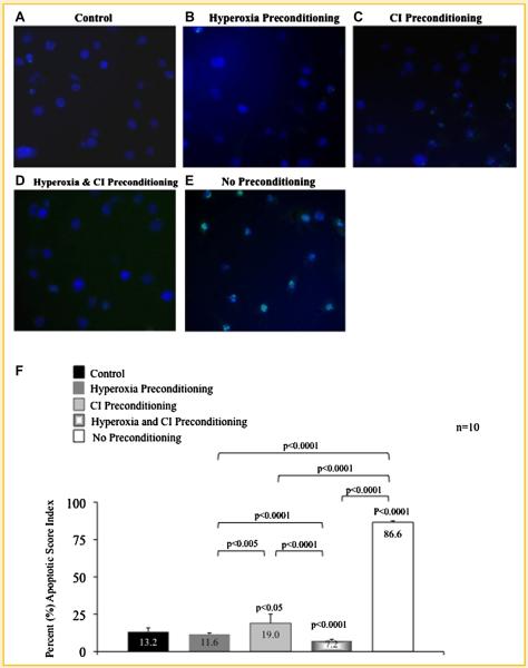
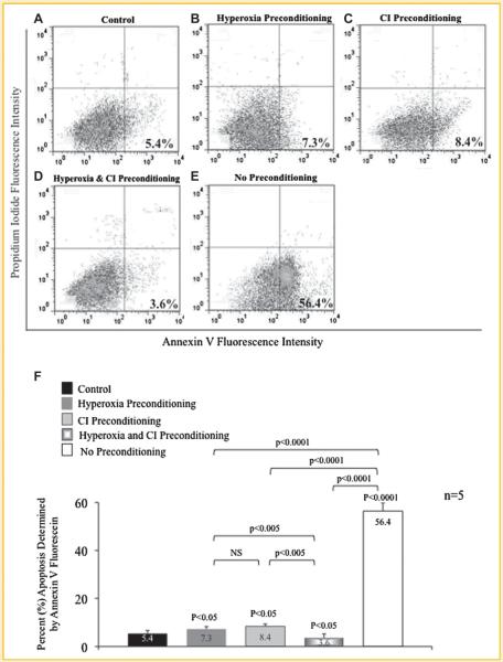
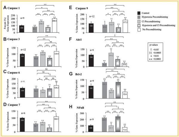
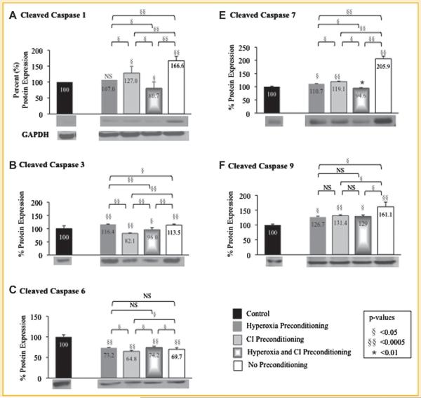
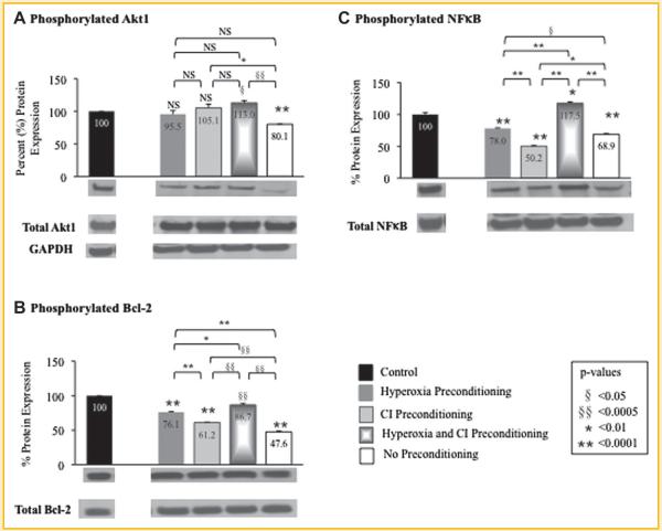
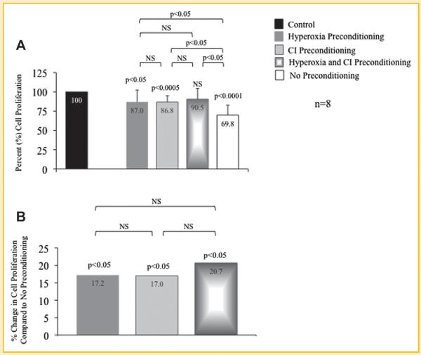
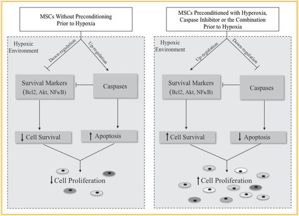
Myocardial infarction is a leading cause of mortality and morbidity worldwide. Occlusion of a coronary artery produces ischemia and myocardial necrosis that leads to left ventricular (LV) remodeling, dysfunction, and heart failure. Stem cell therapy may decrease infarct size and improve LV function; the hypoxic environment, however, following a myocardial infarction may result in apoptosis, which in turn decreases survival of transplanted stem cells. Therefore, the effects of preconditioned mesenchymal stem cells (MSC) with hyperoxia (100% oxygen), Z-VAD-FMK pan-caspase inhibitor (CI), or both in a hypoxic environment in order to mimic conditions seen in cardiac tissue post-myocardial infarction were studied in vitro. MSCs preconditioned with hyperoxia or CI significantly decreased apoptosis as suggested by TUNEL assay and Annexin V analysis using fluorescence assisted cell sorting. These effects were more profound when both, hyperoxia and CI, were used. Additionally, gene and protein expression of caspases 1, 3, 6, 7, and 9 were down-regulated significantly in MSCs preconditioned with hyperoxia, CI, or both, while the survival markers Akt1, NF-κB, and Bcl-2 were significantly increased in preconditioned MSCs. These changes ultimately resulted in a significant increase in MSC proliferation in hypoxic environment as determined by BrdU assays compared to MSCs without preconditioning. These effects may prove to be of great clinical significance when transplanting stem cells into the hypoxic myocardium of post-myocardial infarction patients in order to attenuate LV remodeling and improve LV function.

Keywords: APOPTOSIS; CASPASE INHIBITOR; HYPEROXIA; HYPOXIA; MESENCHYMAL STEM CELLS; PRECONDITIONING.


Figures

Fig. 1
Fig. 1
TUNEL assay of preconditioned mesenchymal stem cells (MSC) following exposure to hypoxia. A: MSCs exposed only to normoxia without treatment (control). B: MSCs preconditioning with hyperoxia; (C) MSCs preconditioned with pan-caspase inhibitor (CI); and (D) MSCs preconditioned with the combination of hyperoxia and CI prior to hypoxia exposure. Preconditioning with hyperoxia, CI, or the combination resulted in a decrease in apoptosis as compared to (E) MSCs exposed to hypoxia without preconditioning. This is obvious by a decrease in green fluorescence which represents apoptotic MSCs. Nucleic DNA has been labeled by DAPI staining (blue fluorescence). F: The apoptotic score index determined by TUNEL assays for MSCs preconditioned with hyperoxia, CI or the combination are shown. P-values above columns represent comparison to control; NS, non- significant.
Fig. 2
Fig. 2
Annexin V expression of preconditioned mesenchymal stem cells (MSC) following exposure to hypoxia. A: MSCs exposed only to normoxia without treatment (control). B: MSCs preconditioned with hyperoxia; (C) MSCs preconditioned with pan-caspase inhibitor (CI); and (D) MSCs preconditioned with the combination of hyperoxia and CI prior to hypoxia exposure. Preconditioning with hyperoxia, CI, or the combination resulted in a decrease in apoptosis as compared to (E) MSCs exposed to hypoxia without preconditioning. This is evident by a decrease in Annexin V expression (percentage shown) quantified by fluorescence assisted cell sorting. Lower right quadrant represents Annexin V positive apoptotic cells; upper right quadrant represents propidium iodide (PI) positive dead cells; and lower left quadrant represents Annexin V and PI negative cells or alive cells (A–E). F: Percent apoptosis of MSCs with and without preconditioning treatment determined by Annexin V expression. P-values above columns represent comparison to control; NS, non-significant.
Fig. 3
Fig. 3
Gene expression of preconditioned mesenchymal stem cells (MSC) following exposure to hypoxia. Quantitative real-time reverse transcription polymerase chain reaction (qRT-PCR) was performed to determine gene expression of caspases (A) 1, (B) 3, (C) 6, (D) 7, and (E) 9, and survival markers (F) Akt1, (G) Bcl-2, and (H) NF-κB in MSCs that underwent preconditioning with hyperoxia, caspase inhibitor (CI), or the combination prior to hypoxia exposure. Gene expression of MSCs exposed to hypoxia is represented as a percent relative to MSCs exposed only to normoxia without treatment (control) set at 100%. MSCs that underwent preconditioning, particularly combination of hyperoxia plus CI, resulted in a decrease in caspases while MSCs that were exposed to hypoxia with no preconditioning had an increase in caspase expression. In addition, preconditioning with hyperoxia or CI resulted in an increase in Akt1, Bcl-2, and NF-κB gene expression while no preconditioning resulted in a decrease in these survival markers after hypoxia. P-values above columns represent comparison to control MSCs; NS, non-significant.
Fig. 4
Fig. 4
Quantitative protein expression and representative Western blots (immediately beneath each graph) of preconditioned mesenchymal stem cells (MSC) following exposure to hypoxia. Western blots were performed to determine protein expression of cleaved caspases (A) 1, (B) 3, (C) 6, (D) 7, and (E) 9 in MSCs that underwent preconditioning with hyperoxia, caspase inhibitor (CI), or the combination prior to hypoxia exposure. Protein expression of MSCs exposed to hypoxia is represented as a percent relative to MSCs exposed only to normoxia without treatment (control) set at 100%. MSCs that underwent preconditioning resulted in a decrease in cleaved caspases compared to MSCs with no preconditioning that were exposed to hypoxia. GAPDH Western blot is also shown as the loading control for all experiments (quantification of protein expression confirmed by computer analysis). P-values above columns represent comparison to control MSCs; NS, non-significant.
Fig. 5
Fig. 5
Quantitative protein expression and representative Western blots (immediately beneath each graph) of preconditioned mesenchymal stem cells (MSC) following exposure to hypoxia. Western blots were performed to determine protein expression of (A) phosphorylated Akt1 at position 473 serine, (B) phosphorylated Bcl-2 at position 70 serine, and (C) phosphorylated NF-κB at position 536 serine in MSCs that underwent preconditioning with hyperoxia, caspase inhibitor (CI), or the combination prior to hypoxia exposure. Protein expression was determined as the ratio of phosphorylated to total protein expression. Phosphorylated protein expression of MSCs exposed to hypoxia is represented on the graphs as a percent relative to MSCs exposed only to normoxia without treatment (control) set at 100%. Preconditioning with hyperoxia and/or CI resulted in an increase in phosphorylated Akt1, Bcl-2, and NF-κB protein expression compared to MSCs with no preconditioning that were exposed to hypoxia. GAPDH Western blot is also shown as the loading control for all experiments (quantification of protein expression confirmed by computer analysis). P-values above columns represent comparison to control MSCs; NS, non-significant.
Fig. 6
Fig. 6
Cell proliferation of preconditioned mesenchymal stem cells (MSC) following exposure to hypoxia. Bromodeoxyuridine (BrdU) assays were performed to determine cell proliferation of MSCs that underwent preconditioning with hyperoxia, pan-caspase inhibitor (CI), or the combination prior to hypoxia exposure. A: Proliferation of MSCs exposed to hypoxia is represented as a percent relative to MSCs exposed only to normoxia without treatment (control) set at 100%. Preconditioned MSCs with the combination of hyperoxia plus CI prior to hypoxia exposure maintained a similar cell proliferation as compared to control MSCs; cell proliferation was the lowest in MSCs exposed to hypoxia without preconditioning. P-values above columns represent comparison to control MSCs. B: MSCs that underwent preconditioning with hyperoxia, CI, or the combination prior to hypoxia exposure resulted in significantly greater cell proliferation compared to MSCs that were exposed to hypoxia without preconditioning.P-values above columns represent comparison to MSCs exposed to hypoxia without preconditioning; NS, non-significant.
Fig. 7
Fig. 7
Schematic diagram showing the effect of mesenchymal stem cells (MSC) preconditioned with hyperoxia, pan-caspase inhibitor, or the combination on apoptosis and cell survival in a hypoxic environment.

References

    1. Abdel-Latif A, Bolli R, Tleyjeh IM, Montori VM, Perin EC, Hornung CA, Zuba-Surma EK, Al-Mallah M, Dawn B. Adult bone marrow-derived cells for cardiac repair a systematic review and meta-analysis. Arch Intern Med. 2007;167:989–997. - PubMed
    1. Assmus B, Honold J, Schachinger V, Britten MB, Fischer-Rasokat U, Lehmann R, Teupe C, Pistorius K, Martin H, Abolmaali ND, Tonn T, Dimmeler S, Zeiher AM. Transcoronary transplantation of progenitor cells after myocardial infarction. N Engl J Med. 2006;355:1222–1232. - PubMed
    1. Bel A, Messas E, Agbulut O, Richard P, Samuel JL, Bruneval P, Hagège AA, Menasché P. Transplantation of autologous fresh bone marrow into infarcted myocardium: A word of caution. Circulation. 2003;108(Suppl.I):II-247–II-252. - PubMed
    1. Boudoulas KD, Hatzopoulos AK. Cardiac repair and regeneration: The Rubik's cube of cell therapy for heart disease. Dis Model Mech. 2009;2:344–358. - PMC - PubMed
    1. Brazil DP, Hemmings BA. Ten years of protein kinase B signaling: A hard Akt to follow. Trends Biochem Sci. 2001;26:657–664. - PubMed

Publication types

MeSH terms