. 2013 Oct;27(10):4089-99.
doi: 10.1096/fj.12-224352. Epub 2013 Jun 24.

The Arp2/3 complex mediates multigeneration dendritic protrusions for efficient 3-dimensional cancer cell migration

Affiliations

The Arp2/3 complex mediates multigeneration dendritic protrusions for efficient 3-dimensional cancer cell migration

Anjil Giri et al. FASEB J. 2013 Oct.

Abstract

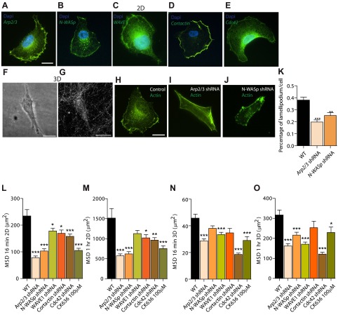
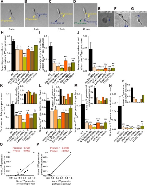
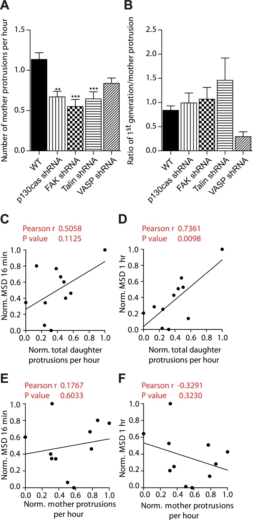
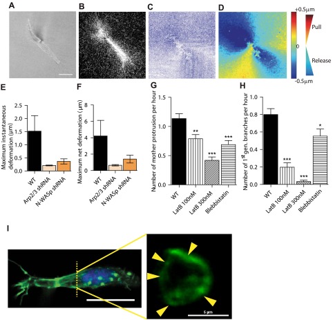
Arp2/3 is a protein complex that nucleates actin filament assembly in the lamellipodium in adherent cells crawling on planar 2-dimensional (2D) substrates. However, in physiopathological situations, cell migration typically occurs within a 3-dimensional (3D) environment, and little is known about the role of Arp2/3 and associated proteins in 3D cell migration. Using time resolved live-cell imaging and HT1080, a fibrosarcoma cell line commonly used to study cell migration, we find that the Arp2/3 complex and associated proteins N-WASP, WAVE1, cortactin, and Cdc42 regulate 3D cell migration. We report that this regulation is caused by formation of multigeneration dendritic protrusions, which mediate traction forces on the surrounding matrix and effective cell migration. The primary protrusions emanating directly from the cell body and prolonging the nucleus forms independent of Arp2/3 and dependent on focal adhesion proteins FAK, talin, and p130Cas. The Arp2/3 complex, N-WASP, WAVE1, cortactin, and Cdc42 regulate the secondary protrusions branching off from the primary protrusions. In 3D matrices, fibrosarcoma cells as well as migrating breast, pancreatic, and prostate cancer cells do not display lamellipodial structures. This study characterizes the unique topology of protrusions made by cells in a 3D matrix and show that these dendritic protrusions play a critical role in 3D cell motility and matrix deformation. The relative contribution of these proteins to 3D migration is significantly different from their role in 2D migration.

Keywords: 3D environment; collagen I matrix; matrix deformation.


Figures

Figure 1.
Figure 1.
Organization and role of Arp2/3 complex and associated molecules in cells on conventional 2D substrates. A–E) Arp2/3 complex (A), N-WASP (B), WAVE1 (C), cortactin (D), and Cdc42 (E) are localized primarily at the leading edge (lamellipodium) of motile cells placed on 2D collagen I-coated substrates. Human fibrosarcoma cells (HT1080) were stained with DAPI (nuclear DNA) and using antibodies against these proteins; images were obtained by immunofluorescence microscopy. F, G) Cells form no apparent wide lamellipodium when embedded in a 3D collagen I matrix; rather they form long pseudopodial protrusions that stem directly from the cell body and branch off into the matrix. Images of the HT1080 cell and its surrounding collagen I matrix were obtained by confocal phase contrast microscopy (F) and confocal reflection microscopy (G), respectively. H–K) Compared to control cells transfected with nontargeting shRNA (H), shRNA-induced depletion of the p34 subunit of the Arp2/3 complex (I), or N-WASP (J) induces the reduction of lamellipodium formation, as measured by the ratio of the length of lamellipodium marked by actin stain (phalloidin) and that of the cell periphery (method described in ref. 44); for each condition, n = 3 and a total of 100 cells were probed (K). **P < 0.01, ***P < 0.001 vs. wild type (WT). L–O) Regulation of 2D (L, M) and 3D (N, O) cell speed (measured as MSD at considered time lags; see Materials and Methods) by the Arp2/3 complex, N-WASP, WAVE1, cortactin, and Cdc42, as well as inhibition following cell treatment by 100 μM of the Arp2/3-complex-specific inhibitor CK636. MSDs were evaluated at time lags of 16 min (L, N), and 1 h (M, O).
Figure 2.
Figure 2.
Cells in matrix form multigeneration, dendritic pseudopodial protrusions. A–D) Dynamic formation of multigeneration, dendritic pseudopodial protrusions by a matrix-embedded cell. Arrows show the formation of a mother protrusion (yellow arrows; protrusions that stem directly from the cell body), as well as first- (blue arrows) and second-generation (green arrows) daughter protrusions, which stem from a mother protrusion. Times: 0 min (A), 8 min (B), 20 min (C), 42 min (D). E–G) Human pancreatic carcinomas (SW1910, E), human prostate carcinomas (E006AA, F), and human epithelial breast carcinomas (MDA-MB-231, G) also form daughter protrusions emanating from mother protrusions when embedded in a 3D collagen I matrix. Scale bars = 20 μm. H–J) Average fraction of time spent by matrix-embedded cells displaying ≥1 first-generation protrusion stemming from a mother protrusion (H), ≥1 second-generation protrusion (I), and ≥1 third-generation protrusion (J), and associated regulation by the Arp2/3 complex, N-WASP, WAVE1, cortactin, and Cdc42, as well as inhibition following cell treatment by 100 μM of the Arp2/3-complex-specific inhibitor CK636. K) Total number of daughter protrusions and mother protrusions (inset) generated per hour per cell (rates of formation). L–N) Number of first-generation protrusions (L), second-generation protrusions (M), and third-generation protrusions (N) generated per hour per cell. Insets: number of first-generation protrusions per mother protrusion (L), number of second-generation protrusions per first-generation protrusion (M), and number of third-generation protrusions per second-generation protrusion (N). O, P) Correlations between the rates of formation of first- and second-generation protrusions (O) and rates of formation of second- and third-generation protrusions (P). For all panels, cells were monitored for 16.5 h. For each condition, n = 3; ≥60 cells were probed. *P < 0.05, **P < 0.01, ***P < 0.001 vs. WT.
Figure 3.
Figure 3.
Daughter protrusions, not mother protrusions, regulate 3D cell speed through the Arp2/3, N-WASP, cortactin, and Cdc42 module. A, B) Total number of mother protrusions produced per hour (A) and degree of branching from mother protrusions (number of first-generation protrusions per mother protrusion; B) in FAK-, talin-, p130Cas-, and VASP-depleted cells. For all panels, cells were monitored for 16.5 h. For each condition, n = 3; ≥60 cells were probed for protrusion analysis. **P < 0.01, ***P < 0.001 vs. WT. C–F) Assessment of correlation between 3D cell speeds evaluated at time lags of 16 min (C, E) and 1 h (D, F) and the rates of formation of daughter protrusions (C, D) and mother protrusions (E, F).
Figure 4.
Figure 4.
Arp2/3 and N-WASP mediate local matrix traction during 3D cell migration and actin architecture of cells. A–D) Particle (PIV) method used to map the time-dependent deformation field of the matrix generated by individual cells in a 3D matrix. Phase contrast (A) and reflection confocal micrograph (B) are simultaneously recorded every 2 min for 2 h to generate instantaneous strain fields of the matrix (C) and determine regions of matrix traction (red; matrix movement toward the cell) and matrix release from the cell (blue; movement away from the cell) (D). E, F) Average instantaneous deformation (E) and maximum deformation (F) of the matrix of fiduciary points located at ∼10 μm distance from the cell. G, H) Rate of formation of mother protrusions (G) and daughter protrusions (H) for control cells and cells treated with actin depolymerizing drug latrunculin B and myosin II inhibitor blebbistatin. *P < 0.05, **P < 0.01, ***P < 0.001 vs. WT. I) Architecture of the actin filament network in matrix-embedded cells, evaluated by confocal microscopy. Maximum confocal projection (left panel) shows the elongated morphology and side protrusions in cells in matrix. Cross sections of the same cell (right panel) reveal that the actin network in protrusions is constituted of cortical bundles. Arrows indicate distinct longitudinal actin filament bundles positioned at the periphery of cell protrusions.

References

    1. Pollard T. D., Borisy G. G. (2003) Cellular motility driven by assembly and disassembly of actin filaments. Cell 112, 453–465 - PubMed
    1. Goley E. D., Welch M. D. (2006) The ARP2/3 complex: an actin nucleator comes of age. Nat. Rev. Mol. Cell Biol. 7, 713–726 - PubMed
    1. Derry J. M., Ochs H. D., Francke U. (1994) Isolation of a novel gene mutated in Wiskott-Aldrich syndrome. Cell 78, 635–644 - PubMed
    1. Miki H., Miura K., Takenawa T. (1996) N-WASP, a novel actin-depolymerizing protein, regulates the cortical cytoskeletal rearrangement in a PIP2-dependent manner downstream of tyrosine kinases. EMBO J. 15, 5326–5335 - PMC - PubMed
    1. Kim A. S., Kakalis L. T., Abdul-Manan N., Liu G. A., Rosen M. K. (2000) Autoinhibition and activation mechanisms of the Wiskott-Aldrich syndrome protein. Nature 404, 151–158 - PubMed

Publication types

MeSH terms

Substances