. 2013 Jul 1;27(13):1435-40.
doi: 10.1101/gad.220202.113. Epub 2013 Jun 24.

An IKKα-E2F1-BMI1 cascade activated by infiltrating B cells controls prostate regeneration and tumor recurrence

Affiliations

An IKKα-E2F1-BMI1 cascade activated by infiltrating B cells controls prostate regeneration and tumor recurrence

Massimo Ammirante et al. Genes Dev. .

Abstract

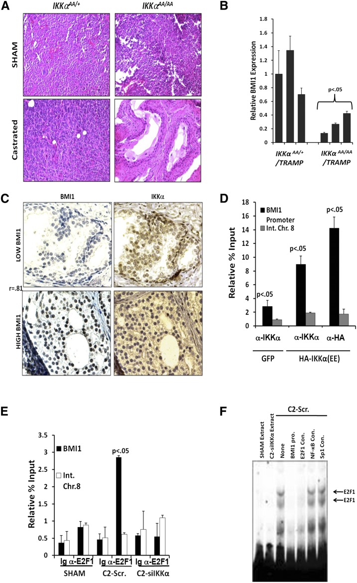
Androgen-deprived prostate cancer (PCa) is infiltrated by B lymphocytes that produce cytokines that activate IκB kinase α (IKKα) to accelerate the emergence of castration-resistant tumors. We now demonstrate that infiltrating B lymphocytes and IKKα are also required for androgen-dependent expansion of epithelial progenitors responsible for prostate regeneration. In these cells and in PCa cells, IKKα phosphorylates transcription factor E2F1 on a site that promotes its nuclear translocation, association with the coactivator CBP, and recruitment to critical genomic targets that include Bmi1, a key regulator of normal and cancerous prostate stem cell renewal. The IKKα-BMI1 pathway is also activated in human PCa.

Keywords: B cells; prostate; regeneration.


Figures

Figure 1.
Figure 1.
B cells and IKKα control prostate regeneration. (A) Prostates of Ikkα+/AA and IkkαAA/AA mice were weighed 17 d after castration and androgen replacement. (B) Prostates of sham-operated or castrated wild-type (WT) mice were collected and analyzed by IHC for IKKα nuclear translocation. Magnification, 400×. Percentages of cells displaying nuclear IKKα are indicated below each panel. n = 3. (C) Prostates of wild-type mice were collected after sham operation or castration and regeneration and analyzed by IF for B220+ cell infiltration. Magnification, 400×. Percentages of B220+ cells are indicated below each panel. n = 3. (D) RNAs extracted from prostates of wild-type mice 17 d after sham operation or castration and subsequent androgen replacement were analyzed for LTα and LTβ expression. (E) Prostates of wild-type, JH−/−, or Cd4−/− mice were weighed 17 d after castration and androgen replacement. In all panels, the values represent mean ± SD; n = 3.
Figure 2.
Figure 2.
IKKα and B cells control prostate and PCa epithelial progenitor proliferation. (A) Single-cell suspensions of Ikkα+/AA and IkkαAA/AA prostates 17 d after castration and androgen replacement were analyzed for sphere-forming ability. After 10 d, the spheres were dissociated, and equal numbers of cells were replated for secondary sphere formation. The number of sphere-forming cells per prostate was calculated by normalizing the observed number of primary and secondary spheres to the total amount of epithelial cells harvested from each prostate. (B,C) Prostates of wild-type (WT) mice collected 17 d after sham operation or castration and subsequent androgen replacement were analyzed by IHC for p63 (B) or BMI1 (C) expression. Percentages of p63+ and BMI1+ cells were determined. n = 3. (D) Sham-operated and regenerated prostates of wild-type mice were analyzed by IF for IKKα (green) and BMI1 (red). Magnification, 200×. (E,F) RNAs extracted from prostates of the indicated genotypes prepared as above were analyzed for Bmi1 mRNA by qRT–PCR. In all panels, the values represent mean ± SD; n = 3.
Figure 3.
Figure 3.
IKKα stimulates E2F1-mediated Bmi1 gene induction. (A) Histological analysis (H&E staining of paraffin-embedded sections; magnification, 100×) of prostates from 24-wk-old Ikkα+/AA/TRAMP and IkkαAA/AA/TRAMP mice that were castrated or sham-operated at 12 wk of age. (B) Prostates of 24-wk-old mice of the indicated genotypes castrated at 12 wk of age were analyzed for Bmi1 mRNA expression by qRT–PCR. (C) Serial sections of paraffin-embedded human PCa tissues were analyzed for nuclear IKKα and BMI1 by IHC. Nuclear staining indices were quantitated, and the correlation coefficient is indicated at the left. Magnification, 400×. (D) ChIP analysis of IKKα recruitment to the Bmi1 promoter. Cross-linked and sheared chromatin from myc-CaP cells stably infected with GFP or IKKα(EE) lentiviruses was immunoprecipitated with IKKα or HA antibodies and analyzed by qPCR with primers to the Bmi1 promoter or an intergenic region of chromosome 8. The percentage input was calculated by determining the ratio of immunoprecipitated DNA with each antibody to a 10% input sample. (E) Chromatin was isolated from myc-CaP tumors established by mock-silenced (Scr.) or IKKα-silenced cells 2 wk after sham operation or castration. After cross-linking and shearing, chromatin was immunoprecipitated with antibodies to E2F1 or control IgG, and the content of Bmi1 promoter DNA was determined as above. (F) Nuclear extracts were prepared from the myc-CaP tumors described above, and E2F1 DNA-binding activity was analyzed by EMSA with the Bmi1 E2F1-binding site. For competition experiments, 100-fold excess of nonradioactive competitor oligonucleotides representing the Bmi1 E2F1 site or consensus binding sites of the indicated transcription factors were added to the reactions 10 min before probe addition. In B, D, and E the values represent mean ± SD; n = 3.
Figure 4.
Figure 4.
IKKα phosphorylates BM1 and controls its nuclear translocation. (A) HEK293T cells were transfected with vectors expressing wild-type (WT) E2F1 or mutants in which the IKKα phosphorylation site was replaced with alanine (A) or glutamate (E) residues. After 48 h, nuclear extracts were prepared and analyzed by immunoblotting. (B) Prostates of wild-type mice were collected after sham operation or castration and androgen replacement and analyzed by IHC for E2F1α. Magnification, 400×. (C) Subcutaneous myc-CaP bearing mock-silenced (Scr.) or IKKα-silenced tumors were collected 2 wk after castration or sham operation and analyzed by IHC for E2F1. Magnification, 400×. (D,E) Chromatin isolated from subcutaneous myc-CaP tumors formed by mock-silenced (Scr.) or IKKα-silenced cells 2 wk after sham operation or castration was subjected to ChIP analysis with antibodies to CBP (D) or Ac-H3 (E). Relative percentage input was determined as above. In D and E, the values represent mean ± SD; n = 3.
Figure 5.
Figure 5.
BMI1 controls castration-resistant tumor growth. (A) myc-CaP tumors were established in wild-type (WT) mice with cells subjected to mock silencing (Scr.), IKKα silencing, or IKKα silencing + ectopic BMI1 expression. When tumors reached 500 mm3, mice were castrated, and tumor volume was measured. (B) RNA from subcutaneous myc-CaP tumors formed by mock-silenced (Scr.) or IKKα-silenced cells was collected 2 or 3 wk after castration and analyzed for Bmi1 expression. (C) RNAs from subcutaneous myc-CaP tumors formed by the cells in A were analyzed for p16 and p19 expression 2 wk after castration. (D) Chromatin from subcutaneous myc-CaP tumors formed by mock-silenced (Scr.) or IKKα-silenced cells collected 2 wk after castration or sham operation was subjected to ChIP analysis using antibodies to BMI1 or control Ig. The presence of Ink4a/Arf sequences was examined by PCR using the primers in Supplemental Figure S13E or the intergenic region from chromosome 8. (E) Subcutaneous myc-CaP tumors formed as in A were analyzed by IHC for ubi-H2A content. (F) Prostates of Ikkα+/AA/TRAMP and IkkαAA/AA/TRAMP mice were collected 12 wk after sham operation or castration and stained for ubi-H2A. (G,H) Paraffin-embedded human prostate sections representing nonmalignant tissue (n = 17) and PCa (n = 13) were stained for BMI1 (G) or ubi-H2A (H). The staining indices are indicated at the bottom. P < 0.01 for the malignant samples. Magnification, 400× for all images. In A–D, the values represent mean ± SD; n = 3.

References

    1. Ammirante M, Luo JL, Grivennikov S, Nedospasov S, Karin M 2010. B-cell-derived lymphotoxin promotes castration-resistant prostate cancer. Nature 464: 302–305 - PMC - PubMed
    1. Beachy PA, Karhadkar SS, Berman DM 2004. Tissue repair and stem cell renewal in carcinogenesis. Nature 432: 324–331 - PubMed
    1. Ben-Neriah Y, Karin M 2011. Inflammation meets cancer, with NF-κB as the matchmaker. Nat Immunol 12: 715–723 - PubMed
    1. Cao Y, Bonizzi G, Seagroves TN, Greten FR, Johnson R, Schmidt EV, Karin M 2001. IKKα provides an essential link between RANK signaling and cyclin D1 expression during mammary gland development. Cell 107: 763–775 - PubMed
    1. Chen J, Trounstine M, Alt FW, Young F, Kurahara C, Loring JF, Huszar D 1993. Immunoglobulin gene rearrangement in B cell deficient mice generated by targeted deletion of the JH locus. Int Immunol 5: 647–656 - PubMed

Publication types

MeSH terms