. 2013 Jun 24;2(6):e53.
doi: 10.1038/oncsis.2013.19.

Tumor-associated soluble uPAR-directed endothelial cell motility and tumor angiogenesis

Affiliations

Tumor-associated soluble uPAR-directed endothelial cell motility and tumor angiogenesis

J S Rao et al. Oncogenesis. .

Abstract

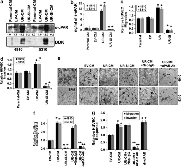
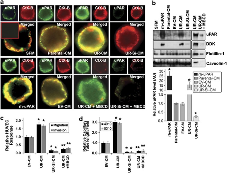
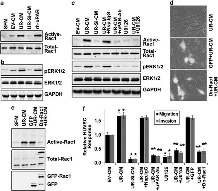
The expression of urokinase-type plasminogen activator (uPA) receptor (uPAR) correlates with the malignant phenotype of various cancers. The soluble form of uPAR (s-uPAR) is present in the circulation of cancer patients, but the role of s-uPAR in endothelial cell migration is poorly understood. Therefore, we examined the role of tumor-associated s-uPAR on endothelial cell motility and angiogenesis. Here, we present evidence that tumor-associated s-uPAR augments the migration of human umbilical vein endothelial cells (HUVECs). When grown on tumor-conditioned medium, the membrane fraction of HUVECs had increased localization of s-uPAR onto its cell membrane. Colocalization studies for GM1 ganglioside receptor and uPAR further demonstrated s-uPAR recruitment onto lipid rafts of HUVECs. Immunoblot analysis for uPAR in lipid raft fractions confirmed s-uPAR recruiting onto HUVECs' membrane. Further, s-uPAR induced Rac1-mediated cell migration while either function-blocking uPAR antibodies or dominant-negative mutant Rac1 expression in HUVECs-mitigated s-uPAR-enhanced cell migration. In addition, orthotopic implantation of uPAR-overexpressing cells resulted in a significant increase in circulating s-uPAR in blood serum and invasive nature of tumor and tumor vasculature in mice. Collectively, this data provide insight into tumor-associated s-uPAR-directed migration of endothelial cells and its subsequent influence on tumor angiogenesis.


Figures

Figure 1
Figure 1
Tumor-associated soluble uPAR (s-uPAR) enhances HUVEC invasion, migration and angiogenesis. (a) Conditioned medium (CM) was collected from tumor cells (parental and stably expressing empty vector (EV), uPAR-cDNA (UR) and uPAR siRNA (UR-Si)). Immunoblot analyses were performed for s-uPAR and DDK using specific antibodies. (b) s-uPAR levels in CM were quantified using uPAR Quantikine Immunoassay kit. Columns: mean; bars: s.d.; n=3; *p<0.01 vs parental control. (c) Cells were labeled (tumor cells: Qtracker-525-Green and HUVECs: Qtracker-655-Red) and seeded into separate chambers of culture inserts. After 16 h, the culture inserts were removed and cells were allowed to migrate for a further 24 h. Images were captured at 0 and 24 h of incubation and cell migration was quantified using ImageJ software (NIH). The levels of HUVEC migration were normalized to HUVEC migration in parental cells and are represented as arbitrary units. Columns: mean; bars: s.d.; n=3; *P<0.01 vs parental control. (d) HUVEC invasion experiments were performed using ThinCertTM inserts as described in Materials and methods. The levels of HUVEC invasion was quantified and normalized to HUVEC invasion in parental-CM. Columns: mean; bars: s.d.; n=3; *P<0.01 vs parental-CM. (e, f) In vitro angiogenesis assay was performed as described in Materials and methods. The degree of angiogenic induction by CM was quantified by ImageJ software (NIH) for the numerical value of the product of the relative capillary length per microscopic field. Serum-free medium (SFM) and recombinant human uPAR (rh-uPAR) in SFM were used as controls (insets). Columns: mean; bars: s.d.; n=3; *P<0.01 vs parental-CM; **p<0.01 vs UR-CM. uPAR antibody, uPAR-Ab; isotype control, NSp.IgG. (g) Migration assay was performed using CM. In this case, both chambers of culture inserts were seeded with HUVECs. After 16 h, the culture inserts were removed, CM was added and cells were allowed to migrate for 24 h. Invasion assay was performed as described above. uPAR-Ab. or Nsp.IgG were added to UR-CM before adding onto cells. rh-uPAR was added to SFM. Columns: mean; bars: s.d.; n=3; *P<0.01 vs parental-CM; **p<0.01 vs UR-CM.
Figure 2
Figure 2
s-uPAR recruits onto HUVEC membrane. Conditioned medium (CM) was collected from tumor cells as described in Materials and methods. (a) HUVECs were cultured on CM for 24 h, labeled with anti-uPAR antibody, followed by Alexa Fluor-488-conjugated secondary antibody and were analyzed by fluorescence-activated cell sorting (FACS) for uPAR expression. Serum-free medium (SFM) and rh-uPAR were used as controls. Isotype control (Neg.). (b) HUVECs were cultured in chamber slides on CM for 24 h and fixed in 4% paraformaldehyde and 0.2% glutaraldedyde in phosphate-buffered saline for 1 h. Immunocytochemical analysis was performed as described in Materials and methods. Isotype control (Neg.; inset). Slides were mounted and photographed. (c) Equal amounts of proteins were used for the extraction of HUVEC membrane fractions and were subjected to immunoblot analysis for uPAR expression using specific antibodies. The blot was re-probed for DDK-tag expression.
Figure 3
Figure 3
s-uPAR colocalizes in lipid rafts on HUVECs. Conditioned medium (CM) was collected from tumor cells as described in Materials and methods. (a) HUVECs were cultured in chamber slides on CM for 24 h and incubated with anti-uPAR antibody followed by Alexa Fluor-488-conjugated secondary antibody at 4 °C. Cells were again labeled with Alexa Fluor-595-CTxB subunit. Slides were mounted and analyzed by confocal microscopy. Negative controls, using an isotype antibody, showed no staining (inset). Serum-free medium (SFM) and DDK-tag containing rh-uPAR were used as controls. To disrupt lipid rafts, HUVECs were pretreated with MBCD, as described in Materials and methods. (b) HUVECs lipid rafts were isolated as described in Materials and methods. Lipid raft-enriched fractions were analyzed for uPAR and DDK-tag levels using immunoblot analysis. Flotillin-1 and caveolin-1 served as controls. Protein band intensities were quantified by densitometric analysis using ImageJ software (NIH). The levels of uPAR protein were normalized to protein levels in HUVECs cultured on parental-CM. Columns: mean; bars: s.d.; n=3; *P<0.01 vs parental-CM. (c) Invasion and migration assays were performed as described in Figure 1d In vitro angiogenesis assay was performed as described in Figure 1. To deplete cholesterol, HUVECs were pretreated with MBCD as described in Materials and methods (c and d). Columns: mean; bars: s.d.; n=3; *p<0.01 vs parental-CM; **p<0.01 vs UR-CM.
Figure 4
Figure 4
s-uPAR induces ERK/Rac1-mediated migration and tube formation in HUVECs. Conditioned medium (CM) was collected from tumor cells, as described in Materials and methods. (a) HUVECs lysates were used to perform GST-Rac1 pull-down assay. The protein complexes were subjected to immunoblot analysis to detect active Rac1. Rac1 from total cell lysates was used as a control. (b) Total cell lysates were subjected to immunoblot analysis for phospho-ERK1/2 (pERK1/2) and total ERK1/2. Glyceraldehyde 3-phosphate dehydrogenase (GAPDH) served as a loading control. HUVECs grown on rh-uPAR were used as a control. (c) HUVECs were cultured on CM alone and/or supplemented with functional blocking anti-uPAR antibody (uPAR-Ab) or isotype control (Nsp.IgG.) or MEK inhibitor (U0126) for 24 h. Cell lysates or GST-Rac1 pull-down protein complexes were subjected to immunoblot analysis to detect active Rac1, Rac1 pERK1/2 and ERK1/2. GAPDH served as a loading control. (d) HUVECs were transfected with dominant-negative mutant Rac1 (Dn-Rac1) for 24 h and cultured on UR-CM. Micrographs were captured for green fluorescent protein (GFP) expression (green) and phase contrast (gray) immediately after the addition of UR-CM (magnification × 60). (e) HUVECs were transfected with Dn-Rac1 for 24 h, cultured on CM for another 24 h, collected and lysed. GST-Rac1 pull-down protein complexes were subjected to immunoblot analysis to detect active Rac1. GFP and Rac1 from total cell lysates were used as controls. (f) HUVECs were transfected with Dn-Rac1 for 24 h and cultured on CM alone and/or supplemented with uPAR-Ab., or Nsp.IgG or U0126 for another 24 h. Invasion and migration assays were performed as described in Figure 1. Columns: mean; bars: s.d.; n=3; *P<0.01 vs parental-CM; **P<0.01 vs UR-CM.
Figure 5
Figure 5
Diverse forms of tumor-associated s-uPAR in vitro and in vivo. (a) Conditioned medium (CM) was collected from tumor cells as described in Materials and methods. CM was subjected to deglycosylation using a deglycosylation kit and analyzed by immunoblot for uPAR using specific antibodies. (b) Equal amount of proteins containing HUVEC lysates were used for extraction of cell membrane fractions and were subjected to deglycosylation, and analyzed by immunoblot for uPAR using specific antibodies. (c) In vivo angiogenic assay was performed by using the dorsal air sac model. 4910EV (EV), 4910UR (UR), 4910UR-Si (UR-Si) cells or a recombinant human uPAR (rh-uPAR) containing chamber was implanted in the dorsal cavity of mice. The micrographs for the presence of tumor-induced neovasculature (microvessels with curved thin structures and many tiny bleeding spots) and pre-existing vasculature (straight) were captured. Representative micrographs are shown. (d, e) Blood was collected from mice orthotopically xenografted with stably expressing EV, UR and UR-Si cells. Total uPAR levels were estimated using a commercial human uPAR Quantikine Immunoassay kit according to the manufacturer's instructions. The data quantification for a set I (n=4; d) and set II (n=6; e), on day 15 and 40, respectively, after cell implantation are shown. Columns: mean; bars: s.d.; *P<0.01 vs parental control. (f) Blood serum (from mice 1–6; on day 40) was subjected to deglycosylation and analyzed by immunoblot for uPAR using specific antibodies. D2-D3, D2-D3 domain containing truncated s-uPAR; D3, D3 domain containing truncated s-uPAR; FL, full-length s-uPAR; .
Figure 6
Figure 6
uPAR overexpression enhances tumor growth, vascularity and s-uPAR recruits onto endothelial cells in vivo. (a) Stably expressing EV, UR and UR-Si cells were injected intracerebrally into mice. Mice were euthanized and brains were collected and fixed as described in Materials and methods. Brain sections were stained with hematoxylin and eosin (H&E) solution, and representative micrographs are shown (upper panel). H&E-stained micrographs showing the tumor invasive front ( × 20; lower panel). (b) Brain tumor areas were calculated using Image Pro Discovery Program software (Media Cybernetics, Inc., Rockville, MD, USA). Columns: mean; bars: s.d.; n=6; *P<0.01 vs parental controls. (c) Immunohistochemical analysis of brain sections using anti-uPAR and anti-vascular endothelial growth factor (VEGF). Blood vessels in tumor sections were visualized with biotin-labeled tomato lectin. Inset: isotype control. (d, e) Fluorescence microscopy for colocalization of an endothelial cell marker (von Willebrand factor (vWF)/anti-CD31) and DDK-tag in tumor sections from mice that were implanted with 4910 EV (EV) and 4910UR (UR) cells. Inset, isotype control.

References

    1. Folkman J. Angiogenesis: an organizing principle for drug discovery. Nat Rev Drug Discov. 2007;6:273–286. - PubMed
    1. Bergers G, Benjamin LE. Tumorigenesis and the angiogenic switch. Nat Rev Cancer. 2003;3:401–410. - PubMed
    1. Lamalice L, Le BF, Huot J. Endothelial cell migration during angiogenesis. Circ Res. 2007;100:782–794. - PubMed
    1. Ossowski L. Effect of antisense inhibition of Urokinase receptor on malignancy. Curr Top Microbiol Immunol. 1996;213:101–112. - PubMed
    1. Bajou K, Noel A, Gerard RD, Masson V, Brunner N, Holst-Hansen C, et al. Absence of host plasminogen activator inhibitor 1 prevents cancer invasion and vascularization. Nat Med. 1998;4:923–928. - PubMed