. 2013 Aug 1;191(3):1164-74.
doi: 10.4049/jimmunol.1203084. Epub 2013 Jun 24.

The role of JAK-3 in regulating TLR-mediated inflammatory cytokine production in innate immune cells

Affiliations

The role of JAK-3 in regulating TLR-mediated inflammatory cytokine production in innate immune cells

Huizhi Wang et al. J Immunol. .

Abstract

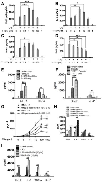
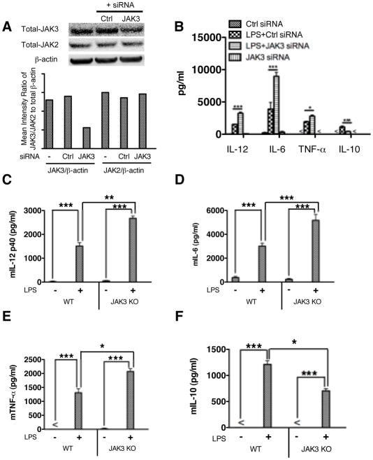
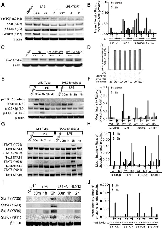
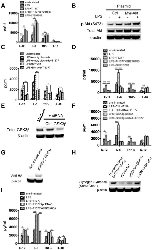
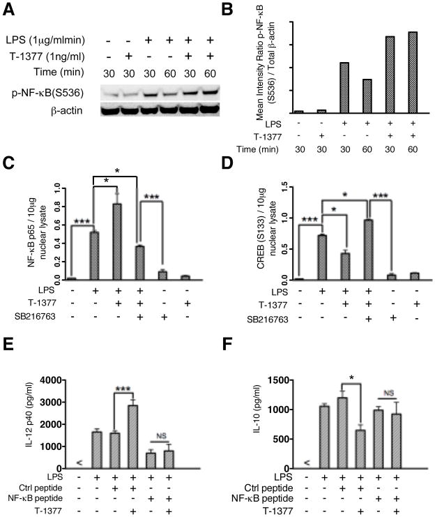
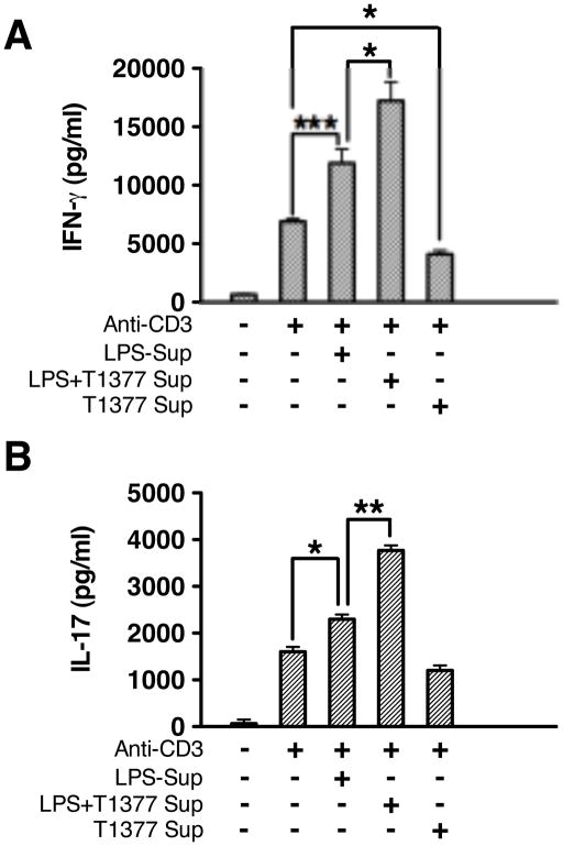
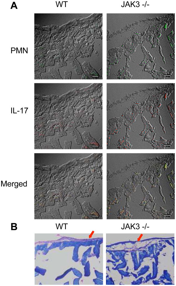
The role of JAK-3 in TLR-mediated innate immune responses is poorly understood, although the suppressive function of JAK3 inhibition in adaptive immune response has been well studied. In this study, we found that JAK3 inhibition enhanced TLR-mediated immune responses by differentially regulating pro- and anti- inflammatory cytokine production in innate immune cells. Specifically, JAK3 inhibition by pharmacological inhibitors or specific small interfering RNA or JAK3 gene knockout resulted in an increase in TLR-mediated production of proinflammatory cytokines while concurrently decreasing the production of IL-10. Inhibition of JAK3 suppressed phosphorylation of PI3K downstream effectors including Akt, mammalian target of rapamycin complex 1, glycogen synthase kinase 3β (GSK3β), and CREB. Constitutive activation of Akt or inhibition of GSK3β abrogated the capability of JAK3 inhibition to enhance proinflammatory cytokines and suppress IL-10 production. In contrast, inhibition of PI3K enhanced this regulatory ability of JAK3 in LPS-stimulated monocytes. At the transcriptional level, JAK3 knockout lead to the increased phosphorylation of STATs that could be attenuated by neutralization of de novo inflammatory cytokines. JAK3 inhibition exhibited a GSK3 activity-dependent ability to enhance phosphorylation levels and DNA binding of NF-κB p65. Moreover, JAK3 inhibition correlated with an increased CD4(+) T cell response. Additionally, higher neutrophil infiltration, IL-17 expression, and intestinal epithelium erosion were observed in JAK3 knockout mice. These findings demonstrate the negative regulatory function of JAK3 and elucidate the signaling pathway by which JAK3 differentially regulates TLR-mediated inflammatory cytokine production in innate immune cells.


Conflict of interest statement

Conflict-of-Interest Disclosure

The authors declare no competing financial interests

Figures

Figure 1
Figure 1. JAK3 inhibitors differentially regulate TLR4-mediated inflammatory cytokine production in human monocytes
Purified human monocytes were pre-treated with different JAK3 inhibitors (T-1377 or WHIP-154 (10μM)) or IL-10 neutralizing antibody (5 μg/ml) using rat IgG1 as an isotype control for 2 hours, and then stimulated with LPS (1μg/ml), Pam3CSK4 (500 ng/ml), or Flagelin (5 μg/ml). After 24 h of stimulation, the cell-free supernatants were collected and the levels of (A) IL-12p40, (B) IL-6, (C) TNF-α, and (D) IL-10 were determined by ELISA. For A–D, lower concentration of T-1377 (<10 nM) enhanced the production of IL-12p40 (A), IL-6 (B), and TNF- α (C) while suppressing IL-10 levels (D) produced by LPS-stimulated human monocytes. JAK3 inhibition by 1 nM T-1377 also differentially regulated production of IL-12 and IL-10 in TLR2- or TLR5-stimulated monocytes (E, F). With different doses of LPS, 1 nM T-1377 exhibited a similar regulatory effect on the production of IL-12 and IL-10 (G). In the context of IL-10 neutralization, JAK3 inhibition (1n M T-1377) remains capable of enhancing LPS-mediated pro-inflammatory cytokine production (H). A second JAK3 inhibitor WHIP-154 (10 μM) showed the same capability to increase or decrease the production of IL-12p40, IL-6, TNF-α and IL-10 (I), respectively. *, **, and *** indicates statistically significant at P<0.05, P<0.01, and P<0.001, respectively. Data represents the arithmetic mean ± S.D. of three biological replicates.
Figure 2
Figure 2. JAK3 deficiency enhances the production of IL-12, TNF-α, and IL-6 while decreasing IL-10 levels in TLR4 stimulated cells
Purified human monocytes were pre-treated with non-target or JAK3 specific siRNA for 72 hours and then stimulated with LPS for 24 h. Whole cell lysates and cell-free supernatants were collected to determine the transfection efficiency and cytokine levels, respectively. (A) siRNA mediated knockdown of JAK3 protein levels was assessed by Western Blot. Total JAK2 levels were also tested to exclude an off-target effect of JAK2. The ratio of total-JAK3 or JAK2 to total β-actin was determined by densitometry. (B) siRNA silencing-mediated JAK3 inhibition enhances the production of IL-12, TNF-α, and IL-6 while decreasing IL-10 levels in LPS stimulated cells. For C to F, wild type and JAK3 knockout BMDM were generated and stimulated with LPS for 24 h. Cell-free supernatants were collected and assayed for cytokine levels by ELISA. JAK3 knockout enhanced TLR4 induced production of IL-12(C), IL-6 (D), TNF-α (E) and reduced IL-10 levels (F) in LPS stimulated BMDM. *, **, and *** indicates statistically significant difference at P<0.05, P<0.01 and P<0.001, respectively. Data represent the arithmetic mean ± S.D. of three biological replicates.
Figure 3
Figure 3. JAK3 inhibition suppresses TLR4-mediated activity of the PI3K pathway while enhancing the phosphorylation levels of STATs
Purified human monocytes were pre-treated with JAK3 inhibitor (1nM T-1377) for 2 h and then stimulated with LPS over a 4 h time course. (A) Total cell lysates were probed for the levels of phosphorylated mTORC1, Akt, GSK3β, and CREB by Western Blot and the ratio of phospho- to total proteins was determined by densitometry (B). (C) Purified human monocytes were stimulated with LPS in the presence or absence of PI3K or GSK3 inhibitor. The levels of phosphorylated JAK3 were analyzed by Western Blot and the ratio to total JAK3 was determined by densitometry (D). From E to H, wild type and JAK3 knockout BMDM were treated with LPS for up to 4 h, total cell lysates were collected at the given time points, and probed for the levels of phosphorylated mTORC1, Akt, GSK3β, CREB (E), STAT3, STAT4, STAT5, and STAT6 (G) by Western Blot and the ratio of phospho- to total protein determined by densitometry (F, H). Anti-IL-12 and anti-IL-6 antibody cocktails were used to determine the effect of JAK3 inhibition on the tyrosine phosphorylation of STAT3, STAT4, STAT5, and STAT6 upon the neutralization of IL-12 and IL-6 signaling. Wild type and JAK3 knockout BMDM were pre-treated with neutralizing anti-IL6 and anti-IL-12 cocktail for 2 h and then stimulated with LPS. Whole cell lysates were probed for the levels of phosphorylated STATs and the ratio of phospho- to total STATs determined by densitometry (I, J). Data are representative of three to five biological replicates.
Figure 4
Figure 4. The ability of JAK3 inhibition to regulate TLR4 mediated inflammatory cytokine production is dependent on the activity of PI3K and GSK3β
The levels of IL-12, TNF-α, IL-6, and IL-10 were assayed by ELISA in LPS stimulated human monocytes. The regulatory effect of JAK3 inhibition was (A) significantly enhanced by PI3K inhibition (LY294002) and (B, C) abrogated by constitutive expression of Akt. Inhibition of GSK3 mediated by pharmacological inhibitor (D) or specific siRNA (E, F) attenuated the regulatory ability of JAK3 on LPS-mediated inflammatory cytokine production in human monocytes. (G) HA expression levels were detected by Western Blot 48 h post-transfection in nontransfected monocytes and monocytes transfected to confirm the transfection efficiency of kinase dead (K85A) plasmid encoding GSK3β. (H) monocytes transfected with kinase dead (K85A) plasmid exhibited a loss in the phosphorylation levels of the GSK3 specific substrate glycogen synthase (GS) (S640/641). (I) As compared with monocytes transfected with empty vector control, the kinase dead (K85A) GSK3β mutant abrogated the ability of JAK3 inhibition to increase the production of TNF-α, IL-12, IL-6, and decrease the IL-10 level in LPS stimulated cells. *, **, and *** indicates statistically significant at P<0.05, P<0.01 and P<0.001, respectively. Data represent the arithmetic mean ± S.D. of three biological replicates.
Figure 5
Figure 5. JAK3 inhibition affects the phosphorylation levels and DNA binding activity of NF-κB and CREB in monocytes
Purified human monocytes were pretreated with JAK3 inhibitor (T-1377, 10 nM), control peptide, or NF-κB inhibitory peptide and then stimulated with LPS. Whole cell lysates, cytosoplasmic and nuclear cell fractions were collected after stimulation with LPS at the time points indicated. (A) JAK3 inhibition increased the phosphorylation of NF-κB (S536) upon LPS stimulation. (B) Densitometer scans of phospho-NF-κB (S536) and total β-actin were performed and recorded as the ratio of phospho-NF-κB (S536): total β-actin. NF-κB and CREB DNA binding assays were performed to measure the levels of total NF-κBp65 (C) (10 μg nuclear lysate) and CREB (S133) (D) (20 μg nuclear lysate) in monocytes stimulated with LPS for 4 h in the presence and absence of the JAK3 inhibitor (T-1377), GSK3 inhibitor (SB216763) or both. For E and F, the functional effect of JAK3 inhibition on NF-κB P65 activity in LPS stimulated monocytes was assessed by pre-treatment of cells with a control peptide or NF-κB inhibitor peptide (100 μM) followed by stimulation with LPS for 20 h; The levels of IL-12P40 (E) and IL-10 (F) were determined by ELISA. For C, D, E, and F, data represents the arithmetic mean ± S.D. of three biological replicates. * and *** indicates statistically significant at P<0.05 and P<0.001, respectively.
Figure 6
Figure 6. Modulation of inflammatory cytokines by JAK3 in human monocytes enhanced Th1 and Th17 cytokine production by memory CD4+ T cells
Human monocytes and autologous memory CD4+ T cells were isolated from PBMC. Monocytes were stimulated with LPS in the presence and absence of JAK3 inhibitor (T-1377) for 24 hours. Cell-free supernatant isolated from non-stimulated monocytes, LPS stimulated monocytes, or monocytes stimulated with LPS in the presence of JAK3 inhibitor were transferred to culture with autologous memory CD4+ T cells plated in 96 well plates (2×10⁵/well) pre-coated with or without anti-CD3 (1 μg/ml). After 96 hours of co-culture, cell-free supernatants were collected and assayed for IFN-γ or IL-17 by ELISA. Data represent the arithmetic mean ± S.D. of three biological replicates * and *** indicates statistically significant at P<0.05 and P<0.001, respectively.
Figure 7
Figure 7. JAK3 knockout aggravated the erosion of intestinal epithelium and enhanced the infiltration of PMN and expression of IL-17
(A) PMN infiltration and IL-17 expression were assessed by staining sections of intestinal tissue with FITC conjugated anti-Ly6G (a specific neutrophil marker) and AlexaFluor 594-conjugated IL-17 (magnification, 40×, scale bar = 50μm). (B) Hematoxylin and Eosin (H & E) staining of serial sections of small intestine from WT mice and JAK3 knockout mice showing the integrity of intestinal epithelium (magnification, 20×)

Similar articles

Cited by

References

    1. O’Neill LA. ‘Fine tuning’ TLR signaling. Nat Immunol. 2008;9:459–461. - PubMed
    1. Foster SL, Hargreaves DC, Medzhitov R. Gene-specific control of inflammation by TLR-induced chromatin modifications. Nature. 2007;447:972–978. - PubMed
    1. Garlanda C, Di Liberto D, Vecchi A, La Manna MP, Buracchi C, Caccamo N, Salerno A, Dieli F, Mantovani A. Damping excessive inflammation and tissue damage in Mycobacterium tuberculosis infection by Toll IL-1 receptor 8/single Ig IL-1-related receptor, a negative regulator of IL-1/TLR signaling. J Immunol. 2007;179:3119–3125. - PubMed
    1. Kawai T, Akira S. TLR signaling. Semin Immunol. 2007;19:24–32. - PubMed
    1. Lee MS, Kim YJ. Signaling pathways downstream of pattern-recognition receptors and their cross talk. Annu Rev Biochem. 2007;76:447–480. - PubMed

Publication types

MeSH terms

Substances