Review
. 2012 Apr;2(2):1303-53.
doi: 10.1002/cphy.c110041.

Pathophysiology of acute kidney injury

Affiliations
Review

Pathophysiology of acute kidney injury

David P Basile et al. Compr Physiol. 2012 Apr.

Abstract

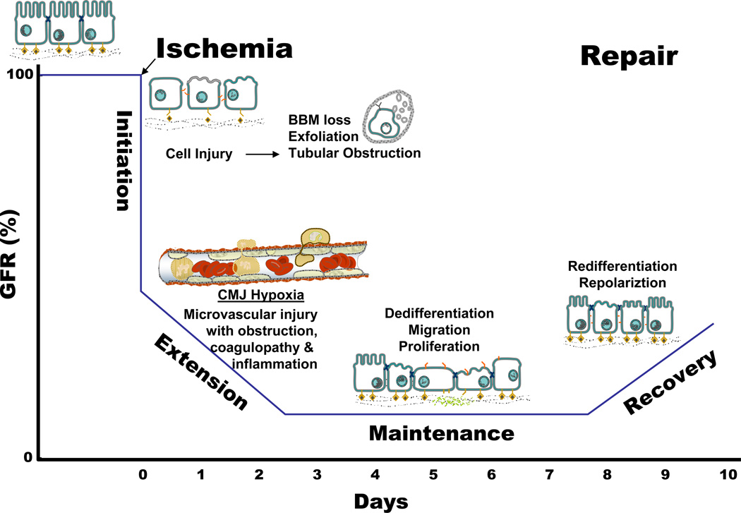
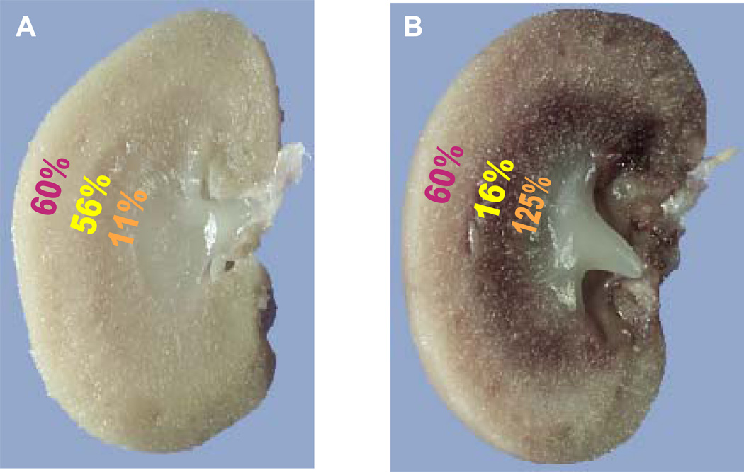
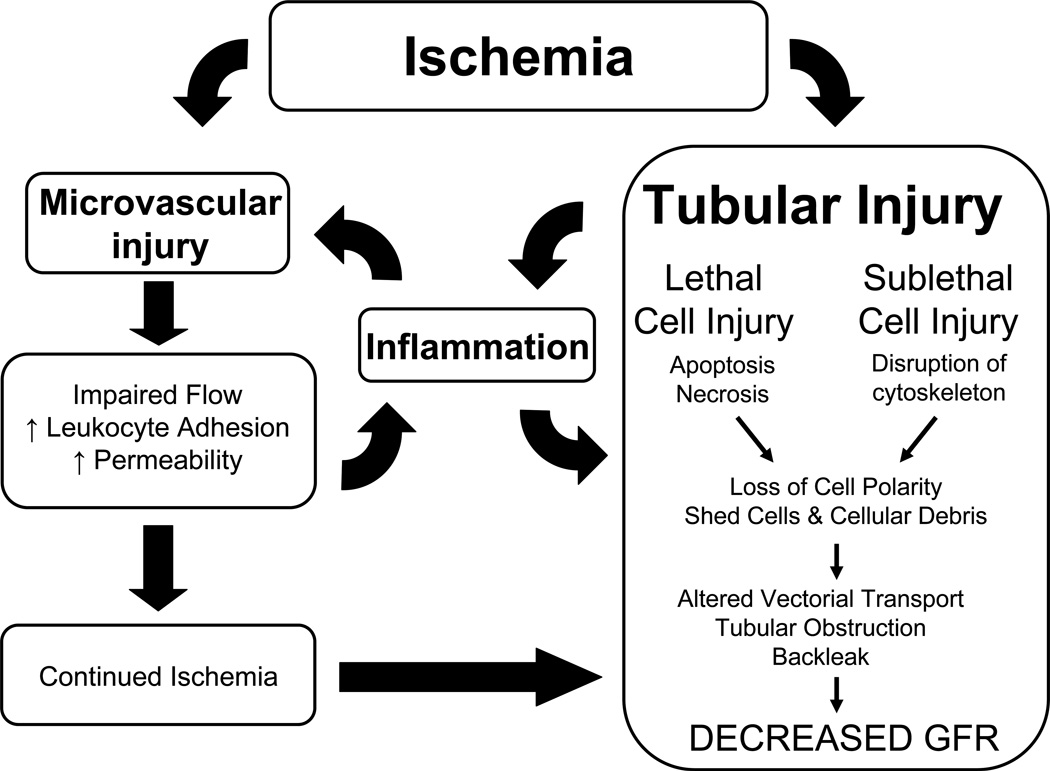
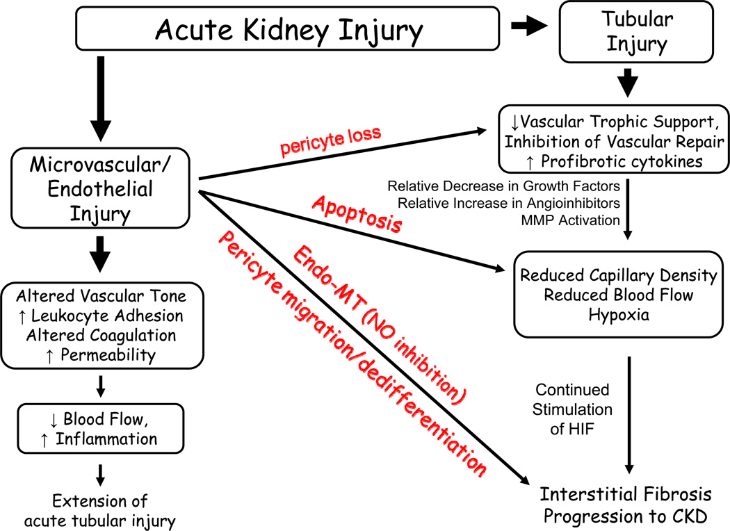
Acute kidney injury (AKI) is the leading cause of nephrology consultation and is associated with high mortality rates. The primary causes of AKI include ischemia, hypoxia, or nephrotoxicity. An underlying feature is a rapid decline in glomerular filtration rate (GFR) usually associated with decreases in renal blood flow. Inflammation represents an important additional component of AKI leading to the extension phase of injury, which may be associated with insensitivity to vasodilator therapy. It is suggested that targeting the extension phase represents an area potential of treatment with the greatest possible impact. The underlying basis of renal injury appears to be impaired energetics of the highly metabolically active nephron segments (i.e., proximal tubules and thick ascending limb) in the renal outer medulla, which can trigger conversion from transient hypoxia to intrinsic renal failure. Injury to kidney cells can be lethal or sublethal. Sublethal injury represents an important component in AKI, as it may profoundly influence GFR and renal blood flow. The nature of the recovery response is mediated by the degree to which sublethal cells can restore normal function and promote regeneration. The successful recovery from AKI depends on the degree to which these repair processes ensue and these may be compromised in elderly or chronic kidney disease (CKD) patients. Recent data suggest that AKI represents a potential link to CKD in surviving patients. Finally, earlier diagnosis of AKI represents an important area in treating patients with AKI that has spawned increased awareness of the potential that biomarkers of AKI may play in the future.


Figures

Figure 1
Figure 1. Relationship between the clinical phases and the cellular phases of ischemic acute kidney injury (AKI), and the temporal impact on organ function as represented by glomerular filtration rate (GFR)
Prerenal azotemia exists when a reduction in renal blood flow causes a reduction in GFR. A variety of cellular and vascular adaptations maintain renal epithelial integrity during this phases. The initiation phase ossuces when a further reduction in renal blood flow results in cellular injury, particularly the renal tubular epithelial cells, and a continued decline in GFR. Vascular and inflammatory processes that contribute to further cell injury and a further decline in GFR usher in the extension phase. During the maintenance phase, GFR reaches a stable nadir as cellular repair processes are initiated in order to maintain and re-establish organ integrity. The recovery phases in marked by a return of normal cell and organ function than results in an improvement in GFR. Adapted from (532).
Figure 2
Figure 2. Regional blood flow is altered following injury in ischemic AKI
Immediately following ischemic injury total renal blood flow is reduced but more striking are the regional deficits in blood flow that exist in the cortex, outer stripe of outer medulla and inner stripe of the outer medulla as indicated in panel A (data from (251)). As overall blood flow starts to recover in the ensuing hours after injury, profound regional alterations in blood flow remain with progressive and profound reduction of the blood flow to the outer stripe of the outer medulla as indicated in B (data from (202)).
Figure 3
Figure 3. Interplay between tubular and vascular injury leading to sustained reductions of GFR in the extension phase of AKI
Injury induced by ischemia can results in damage to both the tubular as well as the microvascular compartment. Resolution of vasoconstriction appears effective at reducing injury when administered prophylactically, but not following established injury. Resistance may be due to exacerbated inflammation, which may impart reductions in RBF and GFR insensitive to vasodilator therapies. Of central importance in this process is the activation of inflammatory processes which are influenced by factors released by damaged proximal tubules as well as adhesion of damaged microvascular cells. Infiltrating leukocytes may impinge on RBF either by secreting vasoactive factors, or by contributing to the disruption of flow by physical interference. In addition, exacerbated hypoxia leading to tubular obstruction may contribute to reductions in GFR independent of vasodilator therapy. From citation (531)
Figure 4
Figure 4. Organization of vascular compartment in the kidney
(A) The medulla is arterial supplied from the efferent arterioles of the juxtamedullary glomeruli, giving supply to the descending arterial vasa recta, and further to the ascending venous vasa recta, draining into the arcuate veins. Abbreviations are: OSOM, outer stripe of outer medulla; ISOM, inner stripe of outer medulla; IM, inner medulla. (B) Very strong expression of b130-1, 2 hours after ischemia/reperfusion (I/R) injury of the kidney, at the level of the ascending vasa recta. (C) Detailed expression of b130-1, 2 hours after I/R injury of the rat kidney, at the level of the ascending venous vasa recta. (D) Detailed expression of b130-1, 2 hours after I/R injury of the human kidney, at the level of the ascending venous vasa recta. (E) Trapping of CD28-expressing T cells in the ascending vasa recta (HIS-17 staining). (F) Trapping of monocytes/macrophages in the ascending vasa recta (ED-1 staining). (G) This trapping of leukocytes in the ascending vasa recta results in an upstream congestion at the ascending arterial vasa recta. This congestion, or no-reflow, represents a well known phenomenon in acute ischemic injury, exacerbating during reperfusion the ischemic damage. From citation (626).
Figure 5
Figure 5. A proposed model for the dephosphorylation, activation, and translocation of ADF to the apical microvilli during ischemia
Under physiological conditions, the distribution of ADF and phosphorylated ADF (pADF) in proximal tubule cells is diffuse throughout the cytoplasm with little or no localization to the apical microvillar region. With ischemia, pADF is dephosphorylated and, therefore, activated. In addition, the diffuse cytoplasmic localization of ADF changes with ADF now concentrating at the apical membrane region of the cell. It is hypothesized that ADF relocalizes to the apical microvillar region and binds the microfilament core, resulting in markedly enhanced filament severing and depolymerization. Breakdown of the microfilament core is accompanied by dramatic changes in the overlying microvillar membrane. The microvillar membrane is internalized or extruded as membrane vesicle or blebs. These vesicles contain both ADF and monomeric actin. Figure and legend from citation (15)
Figure 6
Figure 6. Evidence of mitochondrial depolarization in kidney tissue slices by chemical anoxia using multiphoton imaging
PTs loaded with TMRM showed rapid depolarization of Δψm after chemical anoxia. Bar = 20 µm. (B) In the DT, the decrease was slower and Δψm was not completely depolarized after 60 min of anoxia; however, in the presence of oligomycin (5 µg/ml), Δψm depolarized rapidly in distal tubular cells when exposed to anoxia. Data are means ± SE signal per tubule from a total of 15 PTs, 15 DTs without oligomycin, and 29 DTs with oligomycin from three separate slices for each experiment. The data were normalized from 1 (value at t = 0, taken as resting Δψm) to 0 (minimum value after FCCP, taken as 0 mV). Data from citation (185).
Figure 7
Figure 7. Illustration of the various stages of apoptotic cell death
A: depiction of the stereotypical changes including condensation, changes in nuclear structure, and fragmentation of the cell into small apoptotic bodies. In vivo, the apoptotic bodies are phagocytosed by neighboring cells, whereas in vitro they undergo swelling and eventual lysis (secondary necrosis). B: photographs of LLC-PK1 cells undergoing apoptosis at the corresponding stages as shown in A. Apoptosis was induced by overnight exposure of the cells to 50 µM cisplatin. The cells in the first 3 photographs were stained with Hoechst dye, and the cells in the last photograph were stained with acrydine orange and ethidium bromide. In the last photograph, viable cells appear green, whereas the apoptotic cells with intact plasma membrane appear green with yellowish dots representing condensed chromatin; apoptotic cells and bodies that are undergoing secondary necrosis appear bright orange or red due to the plasma membrane damage and entry of ethidium bromide. Illustration from citation (415).
Figure 8
Figure 8. The continuum of renal cell damage
Individual renal tubular cells are likely to respond in different ways to injury depending upon the severity of the noxious stimulus. The majority of cells presumably remain viable, either because they escape injury altogether, or because they are only sublethally injured and able to recover. More severe injury likely results in apoptosis, whereas necrosis only occurs when cells are subjected to extremely severe injury that leads to critical energy depletion and subsequent metabolic collapse. Legend and figure from citation (320).
Figure 9
Figure 9. Overview of death-signalling pathways in mammalian cells
The death receptor pathway (left) is initiated by the binding of a ligand (Eg: FasL) to its receptor Fas, which results in the sequential recruitment of FADD and pro-caspase-8. c-FLIP can block the recruitment of pro-caspase-8 to the complex. The proximity of several pro-caspase-8 molecules results in its activation. Caspase-8 can proteolytically activate caspase-3, or it can cleave Bid to its truncated form t-Bid, which binds to Bax and gets integrated into the mitochondrial membrane to release cytochrome c. In response to various cellular stress-induced apoptotic stimuli, the intrinsic mitochondrial pathway is activated. This pathway involves the translocation of proapoptotic molecules such as Bax from the cytosol to the mitochondrial membrane. Bax can release cytochromec from the mitochondria into the cytosol. Cytochromec associates with Apaf-1 and caspase-9 to form the apoptosome and subsequent activation of caspase-3. Mitochondria also release AIF and Endo G, which may exert their effects on the nuclei. Mitochondria released Smac/Diablo and Omi/HtrA2 sequesters inhibitors of apoptosis (IAPs) to prevent them from inhibiting caspase-3. BNIP3 is a Bcl2 family member that is translocated and integrated into the mitochondria. Unlike other Bcl2 family members, BNIP3 can induce necrotic cell death in response to death stimuli. Activation of poly (ADP-ribose) polymerase (PARP) leads to NAD+ depletion and may induce mitochondrial depolarization to release AIF. ROS, reactive oxygen species. Legend and Figure from citation (320).
Figure 10
Figure 10. Control of HSP expression in response to cell stress
Shown are the known actions of the constitutively expressed Hsps, primarily of the Hsp70 family called heat shock congnates (HSC) in processing cellular functions. Cell stress increases denatured proteins increasing the demand for HSC. Heat shock transcription factor (HSF), reversibly binds to HSC and is released with the increased demand for HSC. HSF then rapidly initiates transcription for all inducible Hsps including Hsp 70 and Hsp 25/27. Figure from citation (521).
Figure 11
Figure 11. Repair and regeneration of renal proximal tubule cells following acute sublethal injury
Sublethally injured RPTC either repair physiological functions and restore normal tubular function or dedifferentiate, migrate, and/or proliferate to replace lost cells, then differentiate and resume normal function. The processes of repair and regeneration work in concert to ensure relining of the damaged nephron and restoration of renal function. From citation (402).
Figure 12
Figure 12. Evidence that sublethally damaged proximal tubules are the source of dividing cells during recovery from AKI
PAS staining and immunostaining were performed in the kidney sections of creksp;Z/EG mice with renal IRI. (A) PAS staining of the kidney at 2 days after IRI. Tubular injury is shown by the loss of brush border membrane, cell detachment from the basement membrane, and nuclear condensation in some cells (arrows). (B) Expression of PCNA in tubular cells (red, arrows). (C) Low-power image of BrdU incorporation in renal tubules. Some BrdU-containing cells (red, arrows) colocalized with EGFP-expressing cells (green). The arrowhead indicates BrdU incorporation in an EGFP-negative cell. (D) BrdU incorporation (red, arrow) in the epithelial cells expressing EGFP (green). Note: the cre transgene labels tubular cells and their progeny with EGFP. The nuclei were counterstained with DAPI, and images were merged (B–D). Scale bars: 20 µm. From citation (325).
Figure 13
Figure 13. Gross renal morphology and capillary filling in normal and postischemic kidneys
Representative stereoscopic views of 20-µm Microfil-infused kidney section. Shown are Microfil-infused kidneys from a sham-operated rat (A) at 4 (B) and 8 wk (C) post-ischemic injury. In this stereoscopic view, Microfil appears as bright yellow against a dark background. A reduction in Microfil-infused structures in recovered postischemic kidneys is evident. c, cortex; os, outer stripe of the outer medulla; is, inner stripe of the outer medulla; im, inner medulla. Magnification is shown. From citation (32).
Figure 14
Figure 14. A potential role for vascular dropout in promoting the development of CKD following AKI
Acute injury has the potential to affect both tubular and vascular compartments. In addition to direct injury to the microvascular compartment, tubular injury may compromise normal vascular support, shifting the environment to one which promotes vascular impairment rather than vascular stability, including the loss of VEGF expression, the increase in TGF-beta expression as well as several other angio inhibitory compounds. The resultant decrease in capillary structures has a number of potential consquences on renal function including the exacerbation of hypoxia and the impairment of Na handling hemodynamic responses. Hypoxia, along with the potential endothelial mesenchymal transition are likely to participate in the development of fibrosis, which is also influenced by sustained immune/ inflammatory activity. The figure modified from and earlier version published in citation (29).

References

    1. Abul-Ezz SR, Walker PD, Shah SV. Role of glutathione in an animal model of myoglobinuric acute renal failure. Proceedings of the National Academy of Sciences of the United States of America. 1991;88:9833–9837. - PMC - PubMed
    1. Adabag AS, Ishani A, Koneswaran S, Johnson DJ, Kelly RF, Ward HB, McFalls EO, Bloomfield HE, Chandrashekhar Y. Utility of N-acetylcysteine to prevent acute kidney injury after cardiac surgery: A randomized controlled trial. American heart journal. 2008;155:1143–1149. - PubMed
    1. Agarwal A, Balla J, Alam J, Croatt AJ, Nath KA. Induction of heme oxygenase in toxic renal injury: A protective role in cisplatin nephrotoxicity in the rat. Kidney Int. 1995;48:1298–1307. - PubMed
    1. Aki Y, Nishiyama A, Miyatake A, Kimura S, Kohno M, Abe Y. Role of Adenosine A1 Receptor in Angiotensin II- and Norepinephrine-Induced Renal Vasoconstriction. Journal of Pharmacology and Experimental Therapeutics. 2002;303:117–123. - PubMed
    1. Akira S, Takeda K. Toll-like receptor signalling. Nat Rev Immunol. 2004;4:499–511. - PubMed

Publication types

Substances

LinkOut - more resources