. 2013 Jul 9;110(28):11589-94.
doi: 10.1073/pnas.1304365110. Epub 2013 Jun 24.

Optogenetic dissection reveals multiple rhythmogenic modules underlying locomotion

Affiliations

Optogenetic dissection reveals multiple rhythmogenic modules underlying locomotion

Martin Hägglund et al. Proc Natl Acad Sci U S A. .

Abstract

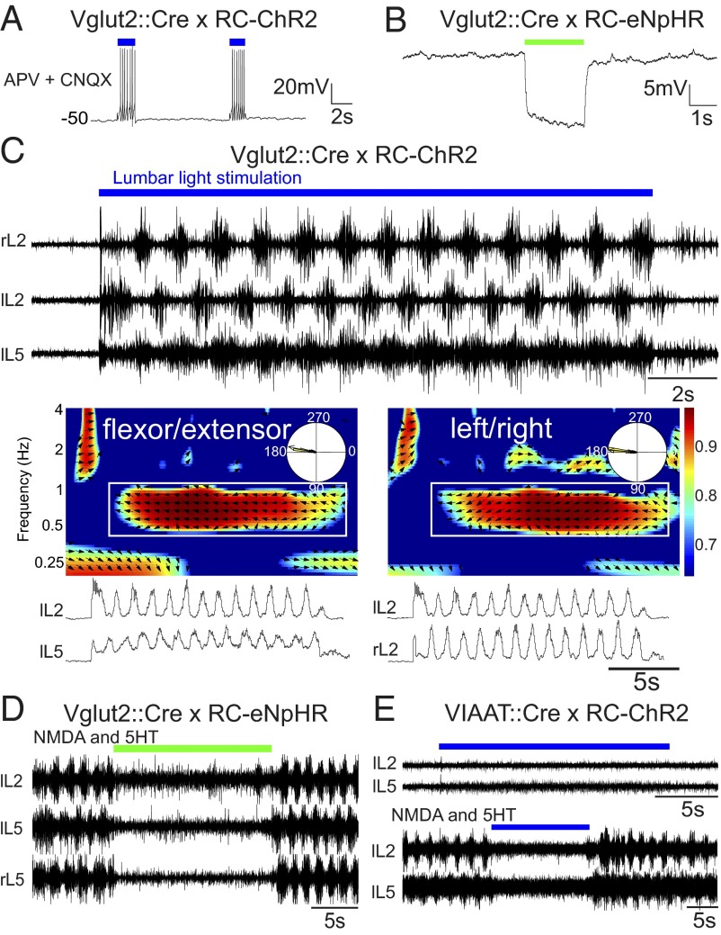
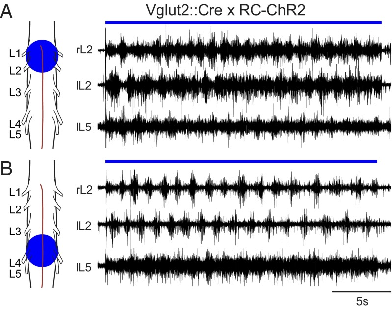
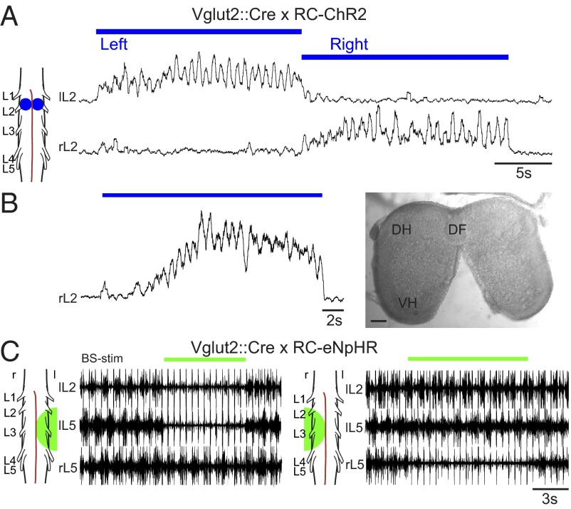
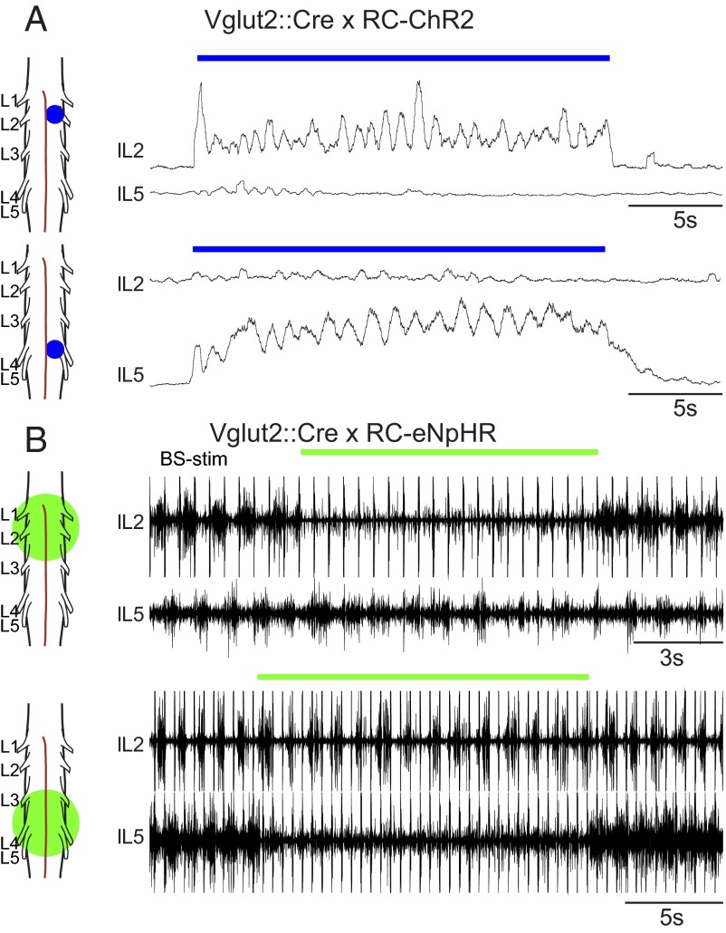
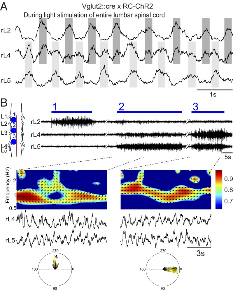
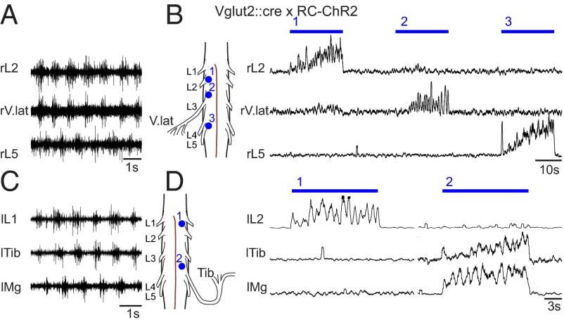
Neural networks in the spinal cord known as central pattern generators produce the sequential activation of muscles needed for locomotion. The overall locomotor network architectures in limbed vertebrates have been much debated, and no consensus exists as to how they are structured. Here, we use optogenetics to dissect the excitatory and inhibitory neuronal populations and probe the organization of the mammalian central pattern generator. We find that locomotor-like rhythmic bursting can be induced unilaterally or independently in flexor or extensor networks. Furthermore, we show that individual flexor motor neuron pools can be recruited into bursting without any activity in other nearby flexor motor neuron pools. Our experiments differentiate among several proposed models for rhythm generation in the vertebrates and show that the basic structure underlying the locomotor network has a distributed organization with many intrinsically rhythmogenic modules.

Keywords: channelrhodopsin-2; halorhodopsin; interneurons; motor neurons.


Conflict of interest statement

The authors declare no conflict of interest.

Figures

Fig. 1.
Fig. 1.
Glutamatergic neurons are both sufficient and necessary for evoking locomotor-like activity, whereas inhibitory neurons can only abolish ongoing locomotor activity. Light depolarizes Vglut2::Cre; RC-ChR2-expressing neurons despite blockade of glutamatergic neurotransmission (AP5 and CNQX) (A) and hyperpolarizes Vglut2::Cre; RC-eNpHR-expressing neurons (B). (C) Stimulating the ventral surface of the lumbar spinal cord of Vglut2::Cre; RC-ChR2 mice evokes locomotor-like activity with direct onset and well-tuned flexor-extensor and left-right coordination. The time-frequency plot shows the coordination between flexor-extensor (Middle Left) and left-right (Middle Right) activity. The color coding denotes the coherence between the two signals and the vector field shows phase delay (also indicated in the circular plots, right = 0°, down = 90°) (Bottom). (D) Inhibiting glutamatergic neurons in the Vglut2::Cre; RC-eNpHR mouse turns off ongoing drug-evoked (5-HT/NMDA: 7 µM/8 µM) activity. Activating inhibitory interneurons does not elicit any rhythmic activity (E) but turns off ongoing drug-evoked (5-HT/NMDA: 7 µM/8 µM) activity.
Fig. 2.
Fig. 2.
Locomotor-like activity can be elicited from rostral or caudal regions. Smaller stimulation areas can evoke locomotor-like activity. Spots ranging from 0.4–2.5 mm2 could successfully evoke locomotor-like activity when centered over rostral (A) or caudal (B) lumbar spinal cord.
Fig. 3.
Fig. 3.
Locomotor-like activity can be initiated and eliminated unilaterally. Stimulating a unilateral spot at L2 in Vglut2::Cre; RC-ChR2 mice elicits unilateral locomotor-like activity in left and right sides consecutively (A), and this activity can be produced when the two sides are separated by cutting the ventral commissure (B; DF, dorsal funiculus; DH, dorsal horn; VH, ventral horn). (Scale bar: 100 µm.) Hyperpolarizing glutamatergic neurons unilaterally, using Vglut2::Cre; RC-eNpHR mice can abolish brainstem-evoked (BS-stim: 2 Hz, 0.5 mA) locomotor-like activity on one side of the cord, whereas activity is maintained on the side contralateral to the light (C; l and r, left and right sides, respectively).
Fig. 4.
Fig. 4.
Flexor and extensor networks can be independently bursting. (A) Activating glutamatergic neurons in a restricted area at the rostral lumbar cord can evoke locomotor-like activity exclusively in the flexor network (Upper), whereas similar activation at more caudal levels evokes locomotor-like activity exclusively in the extensor network (Lower). (B) Inhibiting glutamatergic neurons in the rostral lumbar spinal cord during brainstem evoked locomotor-like activity can turn off the flexor output from the L2 root while the L5 is still rhythmically active (Upper), albeit lower in frequency. Similar inhibition of glutamatergic neurons in the caudal lumbar spinal cord abolishes extensor output from the L5 root and causes a slight reduction in the frequency of the L2 locomotor bursts (Lower).
Fig. 5.
Fig. 5.
The mixed flexor and extensor motor output of L4 can be segregated by site-specific focal light stimulation. The L4 root carries a similar amount of motor output to extensor muscles as to the flexor muscles evident as a double burst pattern seen during locomotor-like activity (by whole preparation Vglut2::Cre x RC-ChR2 stimulation in A). The flexor activity in L2 (B, 1) and L4 (B, 2) can be independently activated depending on the site of focal light stimulation. The L4 output in B2 is flexor-related because it has a phase lag to L5 output (left circle plot). The extensor activity in L4 is also demonstrated by yet another stimulation spot (B, 3 and right circle plot).
Fig. 6.
Fig. 6.
Tibialis anterior or vastus lateralis nerve output can be independently rhythmic without activity in L2. Light simulation of the whole lumbar cord in the Vglut2::Cre; RC-ChR2 mice elicits flexor-related activity in the nerve that projects to vastus lateralis (A) and tibialis anterior (C) muscles. Small spot stimulation shows that these motor neuron pools can be rhythmically active in flexor phase, independent from the rhythmic flexor activity in the L1 and L2 roots (B and D).

Comment in

Similar articles

Cited by

References

    1. Forssberg H (1985) Ontogeny of human locomotor control. I. Infant stepping, supported locomotion and transition to independent locomotion. Exp Brain Res 57(3):480–493. - PubMed
    1. Kudo N, Nishimaru H. Reorganization of locomotor activity during development in the prenatal rat. Ann N Y Acad Sci. 1998;860:306–317. - PubMed
    1. Jordan LM, Liu J, Hedlund PB, Akay T, Pearson KG. Descending command systems for the initiation of locomotion in mammals. Brain Res Brain Res Rev. 2008;57(1):183–191. - PubMed
    1. Lev-Tov A, Etlin A, Blivis D. Sensory-induced activation of pattern generators in the absence of supraspinal control. Ann N Y Acad Sci. 2010;1198:54–62. - PubMed
    1. Kudo N, Yamada T. N-methyl-D,L-aspartate-induced locomotor activity in a spinal cord-hindlimb muscles preparation of the newborn rat studied in vitro. Neurosci Lett. 1987;75(1):43–48. - PubMed

Publication types