. 2013 Jul 9;110(28):11511-6.
doi: 10.1073/pnas.1310144110. Epub 2013 Jun 24.

miR-9 is an essential oncogenic microRNA specifically overexpressed in mixed lineage leukemia-rearranged leukemia

Affiliations

miR-9 is an essential oncogenic microRNA specifically overexpressed in mixed lineage leukemia-rearranged leukemia

Ping Chen et al. Proc Natl Acad Sci U S A. .

Abstract

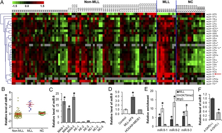
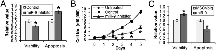
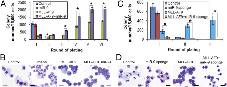
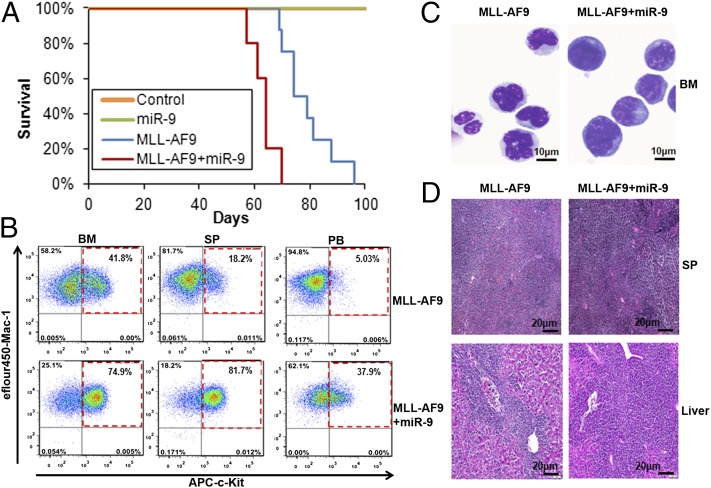
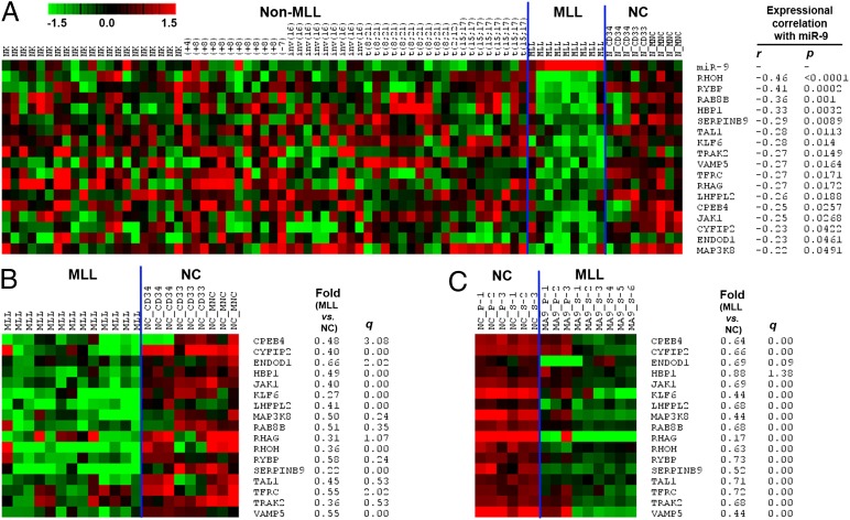
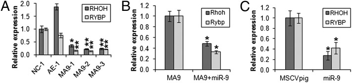
MicroRNAs (miRNAs), small noncoding RNAs that regulate target gene mRNAs, are known to contribute to pathogenesis of cancers. Acute myeloid leukemia (AML) is a group of heterogeneous hematopoietic malignancies with various chromosomal and/or molecular abnormalities. AML with chromosomal translocations involving the mixed lineage leukemia (MLL) gene are usually associated with poor survival. In the present study, through a large-scale, genomewide miRNA expression assay, we show that microRNA-9 (miR-9) is the most specifically up-regulated miRNA in MLL-rearranged AML compared with both normal control and non-MLL-rearranged AML. We demonstrate that miR-9 is a direct target of MLL fusion proteins and can be significantly up-regulated in expression by the latter in human and mouse hematopoietic stem/progenitor cells. Depletion of endogenous miR-9 expression by an appropriate antagomiR can significantly inhibit cell growth/viability and promote apoptosis in human MLL-rearranged AML cells, and the opposite is true when expression of miR-9 is forced. Blocking endogenous miR-9 function by anti-miRNA sponge can significantly inhibit, whereas forced expression of miR-9 can significantly promote, MLL fusion-induced immortalization/transformation of normal mouse bone marrow progenitor cells in vitro. Furthermore, forced expression of miR-9 can significantly promote MLL fusion-mediated leukemogenesis in vivo. In addition, a group of putative target genes of miR-9 exhibited a significant inverse correlation of expression with miR-9 in a series of leukemia sample sets, suggesting that they are potential targets of miR-9 in MLL-rearranged AML. Collectively, our data demonstrate that miR-9 is a critical oncomiR in MLL-rearranged AML and can serve as a potential therapeutic target to treat this dismal disease.


Conflict of interest statement

The authors declare no conflict of interest.

Figures

Fig. 1.
Fig. 1.
MLL fusion proteins directly up-regulate expression of miR-9. (A) Expression profiles of 30 miRNAs that are significantly (q < 0.05; FDR < 0.05; SAM) up-regulated (29 miRNAs) or down-regulated (1 miRNA, i.e., miR-495) in MLL-rearranged AML (n = 10) compared with both normal controls [n = 15, including 6 CD34+ hematopoietic stem/progenitor, 5 CD33+ myeloid progenitor, and 4 MNC cells] and non–MLL-rearranged AML (n = 75). Expression data are mean centered, and the relative value for each sample is represented by a color: high expression is represented with red and low expression is represented with green (scale shown in the upper left). (B) Relative expression levels of miR-9 in 10 human MLL-rearranged AML (i.e., MLL), 75 non–MLL-rearranged AML (i.e., Non-MLL), and 15 normal control (i.e., NC) samples, as detected by Exiqon miRNA microarray assays. (C) qPCR analysis of miR-9 expression level in human cord blood CD34+ cells that were retrovirally transduced with MSCV-MLL-AF9 (MA9-1, -2, and -3), MSCV-AML-ETO (AE-1, -2, and -3), or empty vector (NC-1, -2, and -3). (D) qPCR analysis of miR-9 expression level in colony cells derived from mouse BM progenitor cells retrovirally transduced with MLL-AF9 (MLL-AF9-MSCVneo+MSCVpig), HOXA9+MEIS1 (HOXA9-MSCVpig+MEIS1-MSCVneo), or empty vector (MSCVneo+MSCVpig). (E) ChIP assay of miR-9 promoter regions using anti-MLL (N-terminal), anti-IgG, and anti-H3K79me2 (H3K79 di-methylation) antibodies in MONOMAC-6 cell line. (F) Withdrawal of 4-hydroxy-tamoxifen (4-OHT) (day 7 is shown) results in a significant decrease of miR-9 expression in MLL-ENL-ERtm cell line. *P < 0.05, two-tailed t test.
Fig. 2.
Fig. 2.
Effects of miR-9 on cell viability, proliferation, and apoptosis of human MLL-rearranged AML cells in vitro. (A) Transfection of miR-9 inhibitor (i.e., anti-miR-9 antagomiR oligos) into MONOMAC-6 cells results in a significant decrease of cell viability and an increase in apoptosis compared with transfection of antagomiR scrambled control oligos (as control). (B) Inhibition of miR-9 by miR-9 inhibitor also significantly suppresses cell growth and proliferation of MONOMAC-6 compared with control and untreated MONOMAC-6 cells. Cell numbers were counted daily. (C) Forced expression of miR-9 (via transfection with MSCVpig-miR-9) significantly increases cell viability and decreases apoptosis of MONOMAC-6 cells compared with transfection with MSCVpig empty vector control. Cell viability and apoptosis were evaluated 48 h after transfection. *P < 0.05, two-tailed t test.
Fig. 3.
Fig. 3.
In vitro colony-forming/replating assays. (A) Forced expression of miR-9 promoted MLL-AF9–mediated cell transformation. Normal mouse BM progenitor cells were retrovirally transduced with MSCVneo+MSCVpig (i.e., Control), MSCVneo+MSCVpig-miR-9 (i.e., miR-9), MSCVneo-MLL-AF9+MSCVpig (i.e., MLL-AF9), or MSCVneo-MLL-AF9+MSCVpig-miR-9 (i.e., MLL-AF9+miR-9) and then plated into methylcellulose medium under double selection of puromycin and G418 to form colonies. The colony cells were replated every 7 d for up to six passages. Mean± SD values of colony numbers are shown. (B) Cytospin morphology analysis of first passage of colony cells (see Fig. 3A) via Wright-giemsa staining. (C) Block of miR-9 function inhibited MLL-AF9–mediated cell transformation. Normal mouse BM progenitor cells were retrovirally transduced with MSCVneo+pBABE-puro-scrambled sponge (i.e., Control), MSCVneo+pBABE-puro-miR-9 sponge (i.e., miR-9 sponge), MSCVneo-MLL-AF9+pBABE-puro-scrambled sponge (i.e., MLL-AF9), or MSCVneo-MLL-AF9+pBABE-puro-miR-9 sponge (i.e., MLL-AF9+miR-9 sponge), and colony forming and replating were conducted as described above for up to three passages. Mean ± SD values of colony numbers are shown. (D) Cytospin morphology analysis of first passage of colony cells (see Fig. 3C) via Wright-giemsa staining. The length of bars in B and D represents 10 μm. *P < 0.05; **P < 0.01; two-tailed t test.
Fig. 4.
Fig. 4.
miR-9 promotes MLL-AF9–mediated leukemogenesis in vivo. (A) Kaplan-Meier survival curves of lethally irradiated CD45.1 recipient mice, which were reconstituted with colony cells derived from CD45.2 mouse BM progenitor transduced with MSCVneo+MSCVpig (control, n = 5), MSCVneo+MSCVpig-miR-9 (miR-9; n = 5), MSCVneo-MLL-AF9+MSCVpig (MLL-AF9; n = 8), or MSCVneo-MLL-AF9+MSCVpig-miR-9 (MLL-AF9+miR-9; n = 5). Overexpression of miR-9 accelerates MLL-AF9–mediated leukemia (P = 0.001, log-rank test) compared with the MLL-AF9 alone group. (B) Flow cytometric analysis of bone marrow (BM), spleen (SP), and peripheral blood (PB) from MLL-AF9 and MLL-AF+miR-9 representative mice stained for c-Kit/Mac-1. (C) Wright-Giemsa–stained BM cytospin. (D) H&E-stained SP and liver sections showing massive leukemic infiltration.
Fig. 5.
Fig. 5.
Expression profiles of 17 candidate target genes of miR-9 in MLL-rearranged AML. Expression data were mean centered, and the relative value for each sample is represented by a color, with red representing high expression and green representing low expression (scale shown in the upper left). (A) Expression profiles of miR-9 and its 17 candidate target genes in the 79 human sample set (including 9 MLL-rearranged AML, 61 non–MLL-rearranged AML, and 9 normal control samples) and their expressional correlations are shown. (B) Expression profiles of the 17 candidate target genes in the 22 human sample set including 13 MLL-rearranged AML and 9 normal control samples. (C) Expression profiles of the 17 candidate target genes in the 15 mouse sample set including 9 MLL-rearranged AML and 6 normal control samples. The fold changes and corresponding q values (as detected by SAM) of the 17 target genes in MLL-rearranged AML samples compared with normal controls are shown in B and C.
Fig. 6.
Fig. 6.
RHOH and RYBP are potential direct targets of miR-9. (A) Relative expression of RHOH and RYBP in normal human cord blood CD34+ cells transduced with MSCV-MLL-AF9 (MA9-1, -2, and -3), MSCV-AML1-ETO (AE-1), or empty vector (NC-1) (15). (B) Relative expression of Rhoh and Rybp in normal mouse BM progenitor cells transducted with MSCVneo-MLL-AF9+MSCVpig (MA9) or MSCVneo-MLL-AF9+MSCVpig-miR-9 (MA9+miR-9). Cells used for the analysis are passage II colony cells from the in vitro colony-forming/replating assay shown in Fig. 3A. (C) Relative expression of RHOH and RYBP in human MONOMAC-6 cells 48 h after transfection with empty vector (MSCVpig) or miR-9 (MSCVpig-miR-9). Expression level of each target gene in the control group in each plot was set to 1 for comparison and statistical analysis. *P < 0.05; **P < 0.01; two-tailed t test.

References

    1. He L, Hannon GJ. MicroRNAs: Small RNAs with a big role in gene regulation. Nat Rev Genet. 2004;5(7):522–531. - PubMed
    1. Chen J, Odenike O, Rowley JD. Leukaemogenesis: More than mutant genes. Nat Rev Cancer. 2010;10(1):23–36. - PMC - PubMed
    1. Guo H, Ingolia NT, Weissman JS, Bartel DP. Mammalian microRNAs predominantly act to decrease target mRNA levels. Nature. 2010;466(7308):835–840. - PMC - PubMed
    1. Xiao C, Rajewsky K. MicroRNA control in the immune system: Basic principles. Cell. 2009;136(1):26–36. - PubMed
    1. Löwenberg B, Downing JR, Burnett A. Acute myeloid leukemia. N Engl J Med. 1999;341(14):1051–1062. - PubMed

Publication types

MeSH terms

Associated data

LinkOut - more resources