. 2013 Jul 9;110(28):11296-301.
doi: 10.1073/pnas.1310156110. Epub 2013 Jun 24.

Molecular basis for chromatin binding and regulation of MLL5

Affiliations

Molecular basis for chromatin binding and regulation of MLL5

Muzaffar Ali et al. Proc Natl Acad Sci U S A. .

Abstract

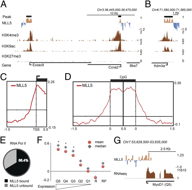
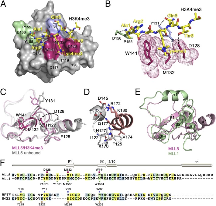
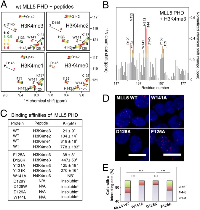
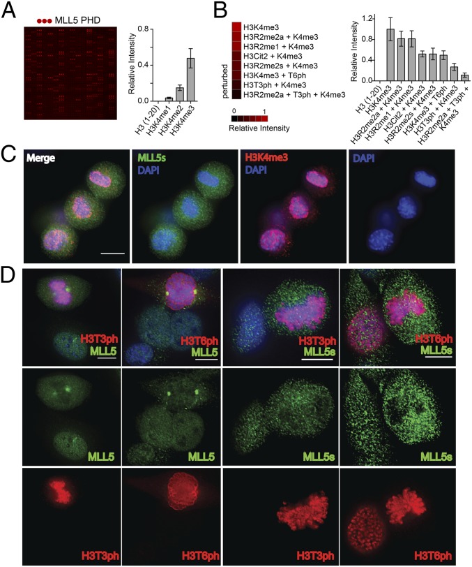
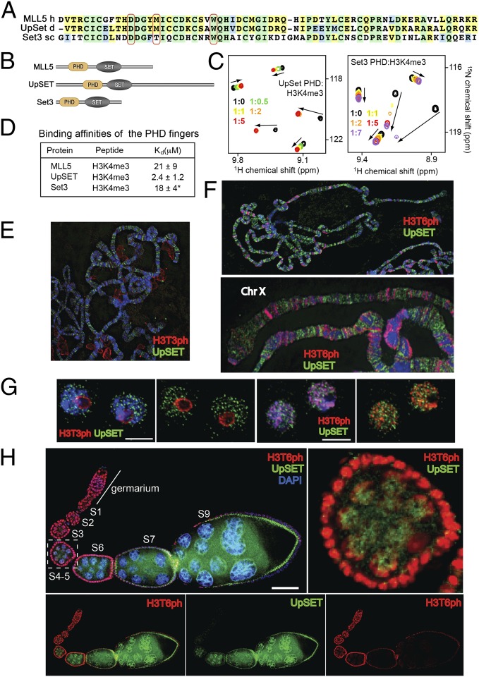
The human mixed-lineage leukemia 5 (MLL5) protein mediates hematopoietic cell homeostasis, cell cycle, and survival; however, the molecular basis underlying MLL5 activities remains unknown. Here, we show that MLL5 is recruited to gene-rich euchromatic regions via the interaction of its plant homeodomain finger with the histone mark H3K4me3. The 1.48-Å resolution crystal structure of MLL5 plant homeodomain in complex with the H3K4me3 peptide reveals a noncanonical binding mechanism, whereby K4me3 is recognized through a single aromatic residue and an aspartate. The binding induces a unique His-Asp swapping rearrangement mediated by a C-terminal α-helix. Phosphorylation of H3T3 and H3T6 abrogates the association with H3K4me3 in vitro and in vivo, releasing MLL5 from chromatin in mitosis. This regulatory switch is conserved in the Drosophila ortholog of MLL5, UpSET, and suggests the developmental control for targeting of H3K4me3. Together, our findings provide first insights into the molecular basis for the recruitment, exclusion, and regulation of MLL5 at chromatin.


Conflict of interest statement

The authors declare no conflict of interest.

Figures

Fig. 1.
Fig. 1.
MLL5 preferentially binds actively transcribed genes. (A and B) MLL5 chromatin profile over a 30-kb region of chromosome 3 (A) or a 5-kb region of chromosome 6 (B). MLL5 DamID signal (a log2 ratio), peaks (a thick black line), and normalized ChIP-seq data for H3K4me3, H3K9ac, and H3K27me3 are shown. The mean signal was smoothed + whiskers. (C) Meta-analysis of MLL5 binding around transcriptional start sites (−1.5 kb upstream; –0.5 bp downstream). (D) Meta-analysis of MLL5 binding around CpG islands (1.5-kb upstream and downstream regions flanking CpG islands). (E) Percentage of MLL5-bound promoter regions overlapping with RNA polymerase II (Table S1). (F) Graph showing the mean and median of MLL5 DamID signal around gene promoters clustered by expression quintiles. High (Q5)- to low (Q1)-expressing gene groups are shown. R, ∼4,000 random sequences; RP, promoter regions randomly selected. Asterisk (*) indicates statistically significant groups in comparison with RP. (G) MLL5 binding over a genomic region containing the MyoD1 gene (quintile 5). Coding regions are shown in black (Fig. S1).
Fig. 2.
Fig. 2.
The crystal structure of the PHD finger of MLL5 in complex with the H3K4me3 peptide. (A) The protein residues involved in the interaction with Lys4me3, Ala1, and Gln5 of the peptide are colored magenta, green, and blue, respectively. (B) A zoom-in view of the H3K4me3 binding pocket. Dashed lines represent intermolecular hydrogen bonds. (C) An overlay of the structure of the H3K4me3-bound MLL5 PHD finger (pink) with the structure of MLL5 PHD in the apo-state (gray; PDB ID code 2LV9). The H3K4me3 peptide is omitted for clarity. (D) A zoom-in view of the MLL5 PHD structure showing close contacts between the N-terminal loop and the C-terminal α-helix. (E) An overlay of the structure of the H3K4me3-bound MLL5 PHD finger (pink) with the structure of the H3K4me3-bound MLL1 PHD3 (green; PDB ID code 3LQJ). (F) Alignment of PHD finger sequences: absolutely, moderately, and weakly conserved residues are colored green, yellow, and blue, respectively. The Lys4me3 binding residues of MLL5, MLL1, BPTF, and ING2 are indicated by magenta, green, blue, and wheat, respectively, squares, and labeled. Secondary structure elements of MLL5 PHD are shown.
Fig. 3.
Fig. 3.
A unique H3K4me3 binding mechanism of MLL5 PHD. (A) Superimposed 1H,15N HSQC spectra of MLL5 PHD collected upon titration of indicated peptides. Spectra are color-coded according to the protein:peptide molar ratio. (B) The normalized chemical-shift changes observed in the PHD finger upon binding to H3K4me3 as a function of residue. (C) Binding affinities of WT and mutated MLL5 PHD for the indicated histone peptides measured by tryptophan fluorescence (a) or NMR (b). NB, no binding. (D and E) HEK293T cells expressing wild-type FLAG-MLL5 and mutants were fixed and stained with anti-FLAG and anti-H3K4me3 antibodies and with secondary antibodies conjugated with Duolink PLA probes and Duolink PLA detection reagents and imaged using a confocal microscope. Foci numbers in each cell were counted manually (400–500 cells were scored for each sample) and grouped into three categories: 1–3, 4–6, and >6. ***P < 0.001.
Fig. 4.
Fig. 4.
Phosphorylation of Thr3 and Thr6 inhibits binding of the MLL5 PHD finger with H3K4me3. (A and B) GST-MLL5 PHD was probed by a histone peptide microarray. Red spots indicate binding of MLL5 PHD to H3K4me3 peptides. The complete list of peptides is shown in Dataset S1. (C) H3K4me3 remains present during mitosis. C2C12 cells were stained with anti-MLL5s (green) and anti-H3K4me3 antibodies (red). Blue indicates DNA stained with DAPI. (D) MLL5 is displaced from chromatin during mitosis. C2C12 cells were stained with MLL5 and MLL5s antibodies (green) and with anti-H3T3ph and anti-H3T6ph antibodies (red). Scale bar, 10 μm.
Fig. 5.
Fig. 5.
Binding to H3K4me3, antagonized by H3T3ph and H3T6ph, is conserved in the MLL5 orthologs Set3 and UpSET. (A) Alignment of the PHD finger sequences. The Lys4me3 binding residues are indicated by red ovals. (B) Architecture of the MLL5/UpSET/Set3 family. (C) Superimposed 1H,15N HSQC spectra of the PHD fingers of UpSET and Set3 collected upon titration of H3K4me3. (D) Binding affinities of the PHD fingers measured by tryptophan fluorescence. (Data from ref. .) (E and F) Third larval instar polytene chromosomes (E) and polytene chromosomes (F) stained with a mix of antibodies to UpSET N- and C-terminus (green), and H3T3ph (red). DNA counterstained with DAPI (blue). See also Fig. S5 A and B. (G) Drosophila S2 cells stained with antibodies to UpSET N terminus (green) and H3T3ph or H3T6ph (red). Scale bar, 5 μm. (H) UpSET protein levels are differentially regulated during oogenesis. Wild-type ovarioles were stained with antibodies against UpSET (green), H3T6ph (red), and costained with DAPI (blue). (Upper Right) Zoom-in of the S4 egg chamber showing a reduced overlap between UpSET and H3T6ph. Scale bar, 100 μm.

References

    1. Emerling BM, et al. MLL5, a homolog of Drosophila trithorax located within a segment of chromosome band 7q22 implicated in myeloid leukemia. Oncogene. 2002;21(31):4849–4854. - PubMed
    1. Heuser M, et al. Loss of MLL5 results in pleiotropic hematopoietic defects, reduced neutrophil immune function, and extreme sensitivity to DNA demethylation. Blood. 2009;113(7):1432–1443. - PubMed
    1. Madan V, et al. Impaired function of primitive hematopoietic cells in mice lacking the mixed-lineage-leukemia homolog MLL5. Blood. 2009;113(7):1444–1454. - PubMed
    1. Zhang Y, et al. MLL5 contributes to hematopoietic stem cell fitness and homeostasis. Blood. 2009;113(7):1455–1463. - PMC - PubMed
    1. Deng LW, Chiu I, Strominger JL. MLL 5 protein forms intranuclear foci, and overexpression inhibits cell cycle progression. Proc Natl Acad Sci USA. 2004;101(3):757–762. - PMC - PubMed

Publication types

Associated data