. 2013 Jul 9;110(28):11302-7.
doi: 10.1073/pnas.1219125110. Epub 2013 Jun 24.

Structural and functional insights into caseinolytic proteases reveal an unprecedented regulation principle of their catalytic triad

Affiliations

Structural and functional insights into caseinolytic proteases reveal an unprecedented regulation principle of their catalytic triad

Evelyn Zeiler et al. Proc Natl Acad Sci U S A. .

Abstract

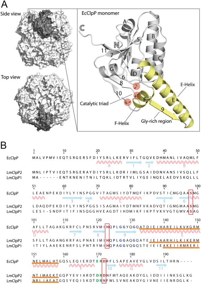
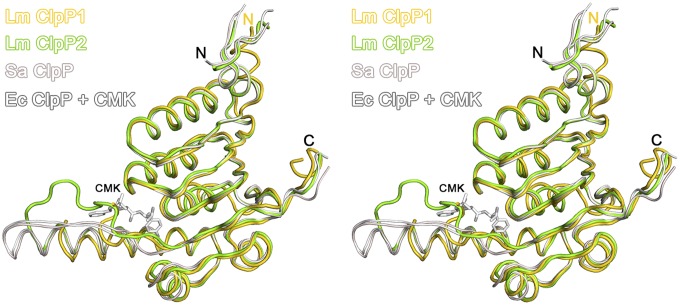
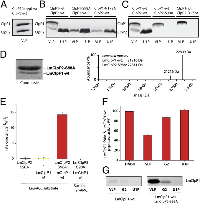
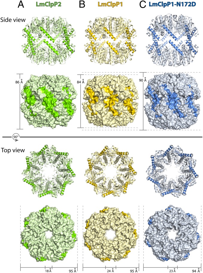
Caseinolytic proteases (ClpPs) are large oligomeric protein complexes that contribute to cell homeostasis as well as virulence regulation in bacteria. Although most organisms possess a single ClpP protein, some organisms encode two or more ClpP isoforms. Here, we elucidated the crystal structures of ClpP1 and ClpP2 from pathogenic Listeria monocytogenes and observe an unprecedented regulation principle by the catalytic triad. Whereas L. monocytogenes (Lm)ClpP2 is both structurally and functionally similar to previously studied tetradecameric ClpP proteins from Escherichia coli and Staphylococcus aureus, heptameric LmClpP1 features an asparagine in its catalytic triad. Mutation of this asparagine to aspartate increased the reactivity of the active site and led to the assembly of a tetradecameric complex. We analyzed the heterooligomeric complex of LmClpP1 and LmClpP2 via coexpression and subsequent labeling studies with natural product-derived probes. Notably, the LmClpP1 peptidase activity is stimulated 75-fold in the complex providing insights into heterooligomerization as a regulatory mechanism. Collectively, our data point toward different preferences for substrates and inhibitors of the two ClpP enzymes and highlight their structural and functional characteristics.


Conflict of interest statement

Conflict of interest statement: M. Gersch, M.P., M.D., and S.A.S. are named inventors on a patent application describing fluorogenic substrates suitable for ClpP activity measurements.

Figures

Fig. 1.
Fig. 1.
Main structural elements of ClpP. (A) Top and side view of the tetradecameric ClpP complex from E. coli (10) (EcClpP, PDB ID code 1TYF, surface representation) with one subunit highlighted in dark gray. Each subunit (close-up, ribbon diagram) is made up of seven α-helices (denoted with letters) and 11 β-strands (denoted with numbers) and contains a catalytic triad (highlighted with red circles). Relevant secondary structures (α-helices E and F, β-strand 9) are highlighted in gold. (B) Sequence alignment of EcClpP with LmClpP1 and LmClpP2. The secondary structure elements are depicted for EcClpP. The catalytic triad is framed in red, the residues forming the E-helix are underlined in orange, the conserved proline and the glycins in the Gly-rich loop are colored blue, and the Asp/Arg sensor is shown in green.
Fig. 2.
Fig. 2.
Stereo-representation of ClpP monomers. Structural superposition of LmClpP1 (gold), LmClpP2 (green), SaClpP (PDB ID code 3V5E, pale red), and EcClpP (PDB ID code 2FZS, gray) with covalently bound CMK inhibitor.
Fig. 3.
Fig. 3.
Activity based protein profiling experiments with β-lactone probes and peptidase activity of different LmClpP constructs. (A) Schematic illustration of a labeling experiment with a ClpP subunit containing the Ser–His–Asp catalytic triad. For R1 and R2, please refer to Fig. S3A. (B) Fluorescent SDS/PAGE analysis of LmClpP1 separately as wild-type and with mutated N172 residue, labeled with VLP and the monocyclic β-lactones (VLP, G2, E2, U1P) in situ. (C) Peptidase activity of LmClpP proteins (3 µM) measured with 200 µM Suc–Leu–Tyr–AMC at 32 °C. (D) Peptidase activity of LmClpP proteins (3 µM) recorded with 100 µM of a custom-synthesized, fluorogenic Leu–ACC (for structural details on the fluorogenic substrate Leu–ACC, see Fig. S5A) substrate at 32 °C. Note, LmClpP1–N172D is 20-fold more active than wild-type LmClpP1. (E) Fluorescent SDS/PAGE analysis of ClpP1 and ClpP1–N172D in comparison with ClpP2, labeled in situ with VLP and monocyclic β-lactones (VLP, U1P, E2, G2, D3, 120P, A1, M1, N1, P1, Q1).
Fig. 4.
Fig. 4.
ClpP1P2 heteroconstructs illustrating distinct substrate specificities. (A) Labeling profile of the isolated tetradecameric complex of LmClpP1(strep)/LmClpP2 with VLP. (B and C) Fluorescent SDS/PAGE analysis of recombinantely coexpressed ClpP1P2 either with ClpP2 wild-type (wt) and a ClpP1 mutant (B) or ClpP2 wild-type and a mutated ClpP1 (C). The labeling was performed with VLP and the monocyclic β-lactone U1P. (D) Copurified Strep-tagged LmClpP2–S98A and tag-free LmClpP1 (ratio 5:1) identified by intact protein MS. (E) Heterooligomerization of LmClpP1 stimulates its peptidolytic activity 75-fold. Rate constants of the heterooligomeric complex were calculated with respect to the concentration of the LmClpP1 subunits. (F) Peptidase activities of the heterooligomeric complex after incubation with 50 µM of inhibitor at room temperature. (G) Binding of isolated LmClpP1 and in the presence of LmClpP2–S98A with β-lactones (VLP, G2, U1P).
Fig. 5.
Fig. 5.
Structure of LmClpP tetradecamers. Top and side view of the tetradecameric complex LmClpP2 (A, green), LmClpP1 (B, gold), and LmClpP1–N172D (C, blue). The E-helix is highlighted with a dark color.
Fig. 6.
Fig. 6.
Mechanism that links activity to oligomerization. (A) Tetradecameric complex of LmClpP1–N172D (blue) structurally superimposed with wt (gold, only two opposite monomers are shown). The monomers superposition of ClpP1 wt (gold) and ClpP1–N172D (blue) show the key elements involved in the catalytic triad. When the active site is aligned in the case of LmClpP1–N172D, proline 125 induces a conformational switch toward the E-helix (A140–T158), which results in the extension of the E-helix (I136–T158) and thus in an extended complex. Furthermore, the alignment of the triad induces the down position of the Asp/Arg (D170/R171) sensor. (B) The close-up displays the Asp/Arg sensor (D170/R171) and the interaction to the sensor of the adjacent monomer of the opposite ring (D′170/R′171), which is absent in wt ClpP1.
Fig. 7.
Fig. 7.
Extension of both heptameric rings in the ClpP tetradecamer. Depicted is a model displaying all key residues within the inactive (compressed) and active (extended) tetradecamer. Activation proceeds through an extension of the E-helix, proline rearrangement, active site alignment, and interaction of the Asp/Arg residues across the heptamer interface.

References

    1. Gottesman S, Roche E, Zhou Y, Sauer RT. The ClpXP and ClpAP proteases degrade proteins with carboxy-terminal peptide tails added by the SsrA-tagging system. Genes Dev. 1998;12(9):1338–1347. - PMC - PubMed
    1. Gottesman S, Maurizi MR, Wickner S. Regulatory subunits of energy-dependent proteases. Cell. 1997;91(4):435–438. - PubMed
    1. Gottesman S, Maurizi MR. Regulation by proteolysis: Energy-dependent proteases and their targets. Microbiol Rev. 1992;56(4):592–621. - PMC - PubMed
    1. Frees D, Qazi SN, Hill PJ, Ingmer H. Alternative roles of ClpX and ClpP in Staphylococcus aureus stress tolerance and virulence. Mol Microbiol. 2003;48(6):1565–1578. - PubMed
    1. Frees D, Sørensen K, Ingmer H. Global virulence regulation in Staphylococcus aureus: Pinpointing the roles of ClpP and ClpX in the sar/agr regulatory network. Infect Immun. 2005;73(12):8100–8108. - PMC - PubMed

Publication types

LinkOut - more resources