. 2013 Jul 9;110(28):11314-9.
doi: 10.1073/pnas.1306768110. Epub 2013 Jun 24.

Structural basis of cargo recognitions for class V myosins

Affiliations

Structural basis of cargo recognitions for class V myosins

Zhiyi Wei et al. Proc Natl Acad Sci U S A. .

Abstract

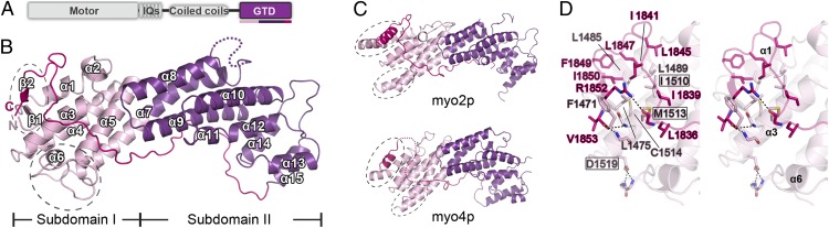
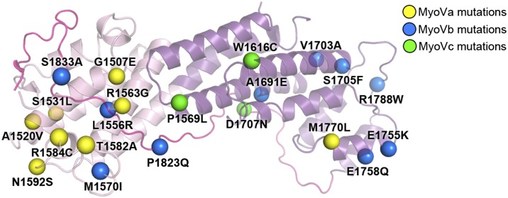
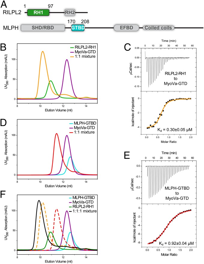
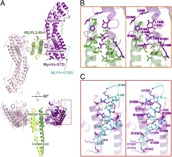
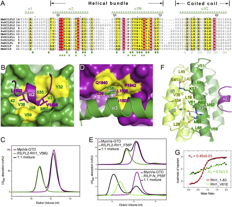
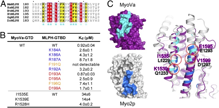
Class V myosins (MyoV), the most studied unconventional myosins, recognize numerous cargos mainly via the motor's globular tail domain (GTD). Little is known regarding how MyoV-GTD recognizes such a diverse array of cargos specifically. Here, we solved the crystal structures of MyoVa-GTD in its apo-form and in complex with two distinct cargos, melanophilin and Rab interacting lysosomal protein-like 2. The apo-MyoVa-GTD structure indicates that most mutations found in patients with Griscelli syndrome, microvillus inclusion disease, or cancers or in "dilute" rodents likely impair the folding of GTD. The MyoVa-GTD/cargo complex structure reveals two distinct cargo-binding surfaces, one primarily via charge-charge interaction and the other mainly via hydrophobic interactions. Structural and biochemical analysis reveal the specific cargo-binding specificities of various isoforms of mammalian MyoV as well as very different cargo recognition mechanisms of MyoV between yeast and higher eukaryotes. The MyoVa-GTD structures resolved here provide a framework for future functional studies of vertebrate class V myosins.

Keywords: MLPH; RILPL2; granuphilin; myo5a; myosin V.


Conflict of interest statement

The authors declare no conflict of interest.

Figures

Fig. 1.
Fig. 1.
Structural characterization of cargo-free MyoVa-GTD. (A) The domain organizations of class V myosins. The color coding of the regions is applied in other panels of this figure. (B) Ribbon representation of the MyoVa-GTD structure. The disordered loop connecting α7 and α8 is indicated by a dotted line. (C) Structural comparisons of MyoVa-GTD with GTD of myo2p (Protein Data Base code 2F6H) and myo4p (3MMI). The N terminus, CC-loop interface, and the α5/α7 connecting region in MyoVa in B and corresponding regions in myo2p and myo4p are highlighted by dashed circles. (D) The molecular details of the N terminus and the CC-loop interface of MyoVa-GTD showing the tight packing via extensive hydrophobic interactions. I1510, M1513, and D1519 are within or close to the interface; mutations of these residues cause a dilute phenotype in mice.
Fig. 2.
Fig. 2.
Disease-causing missense mutations of MyoV-GTD. The mutation sites (indicated by colored spheres) are mapped to the MyoVa-GTD structure (see Table S2 for the list of the mutations).
Fig. 3.
Fig. 3.
Biochemical characterizations of the interactions between MyoVa-GTD and its two cargos, RILPL2 and MLPH. (A) The domain diagrams of RILPL2 and MLPH. SHD, synaptotagmin-like protein homology domain; RBD, Rab-binding domain. The boundaries of the proteins used for the binding assays are indicated. (B, D, and F) Analytical gel filtration chromatography analysis of the MyoVa-GTD/cargo interactions. The profiles of the 1:1 mixtures of MyoVa-GTD/RILPL2-RH1 and MyoVa-GTD/MLPH-GTBD overlap in F as dashed curves. (C and E) The ITC curves showing the quantitative binding affinities between MyoVa-GTD and the two cargos.
Fig. 4.
Fig. 4.
The MyoVa-GTD/RILPL2-RH1/MLPH-GTBD complex structure. (A) Ribbon representation of the 2:2:2 hexamer structure of the MyoVa-GTD/RILPL2-RH1/MLPH-GTBD complex. MyoVa-GTD, RILPL2-RH1, and MLPH-GTBD in one 1:1:1 trimer are colored in purple, green, and cyan, with those in the other identical trimer colored in corresponding lighter colors. This color-coding scheme is used hereafter except as otherwise indicated. (B and C) The detailed interactions between MyoVa-GTD and RILPL2-RH1 (B) and between MyoVa-GTD and MLPH-GTBD (C) in the corresponding regions boxed in A. The disordered N- and C-termini of MLPH-GTBD are indicated by dotted lines. Hydrogen bonds and salt bridges are indicated by dashed lines.
Fig. 5.
Fig. 5.
Characterizations of the MyoVa-GTD/RILPL2-RH1 interaction. (A) The sequence alignment of the RH1 regions of the RILP family members. Dm, Drosophila melanogaster; Dr, Danio rerio; Hs, human; Mm, mouse; Xt, Xenopus tropicalis. Residues that are identical and highly similar are shown in red and yellow boxes, respectively. The secondary structural elements of RILPL2-RH1 are labeled above the alignment. The residues forming RILPL2 dimer interfaces in the helical bundle and coiled-coil regions are indicated by solid and open circles, respectively. The residues involved in the MyoVa-GTD/RILPL2-RH1 interaction are indicated by solid stars. (B and D) The MyoVa-GTD/RILPL2-RH1 interaction is mainly mediated by hydrophobic interactions. Two hydrophobic residues in α2MyoVa, L1502 and V1498, interact with the hydrophobic pocket of RILPL2-RH1 (B). Meanwhile, F56 in α3RILPL2 inserts its aromatic ring into the hydrophobic pocket of MyoVa (D). (C and E) Two sets of analytical gel filtration analysis were used for verification of the structural findings shown in B and D, respectively. (F) The structural basis of the four-helix, bundle-mediated dimer formation of RILPL2. α2MyoVa was shown to indicate the MyoVa binding site on RILPL2. (G) ITC-based analysis showing that truncation of the coiled-coil of RILPL2-RH1 (RH1_1-83) did not affect the MyoVa-GTD/RILPL2-RH1 interaction, whereas disruption of the RILPL2 homodimer (RH1_V61E) largely diminished this interaction.
Fig. 6.
Fig. 6.
Characterizations of the MyoVa-GTD/MLPH-GTBD interaction. (A) The multisequence alignment of MLPH-GTBD. Bt, bovine; Gg, chicken. The residues involved in the MyoVa-GTD/MLPH-GTBD interaction are indicated by solid stars. The negatively charged residues in the MLPH-GTBD C terminus may be involved in the charge–charge interaction with K1540, K1543, and K1544 (Fig. 4C) and are indicated by open stars. (B) The ITC-derived dissociation constants of the bindings between MyoVa-GTD and MLPH-GTBD. (C) Structural comparison of the MLPH-GTBD binding surface (cyan) on MyoVa-GTD and the Vac17/Mmr1-binding surface (blue) on myo2p-GTD. The four overlapped binding residues in MyoVa (purple) and myo2p (gray) are labeled.

References

    1. Johnston GC, Prendergast JA, Singer RA. The Saccharomyces cerevisiae MYO2 gene encodes an essential myosin for vectorial transport of vesicles. J Cell Biol. 1991;113(3):539–551. - PMC - PubMed
    1. Mercer JA, Seperack PK, Strobel MC, Copeland NG, Jenkins NA. Novel myosin heavy chain encoded by murine dilute coat colour locus. Nature. 1991;349(6311):709–713. - PubMed
    1. Espreafico EM, et al. Primary structure and cellular localization of chicken brain myosin-V (p190), an unconventional myosin with calmodulin light chains. J Cell Biol. 1992;119(6):1541–1557. - PMC - PubMed
    1. Hammer JA, 3rd, Sellers JR. Walking to work: Roles for class V myosins as cargo transporters. Nat Rev Mol Cell Biol. 2012;13(1):13–26. - PubMed
    1. Sellers JR, Weisman LS. Myosin V. In: Coluccio LM, editor. Myosins, a Superfamily of Molecular Motors. Vol 9. Dordrecht: Springer; 2008. pp. 289–323.

Publication types