. 2013 Sep;81(9):3264-75.
doi: 10.1128/IAI.00268-13. Epub 2013 Jun 24.

Altered innate defenses in the neonatal gastrointestinal tract in response to colonization by neuropathogenic Escherichia coli

Affiliations

Altered innate defenses in the neonatal gastrointestinal tract in response to colonization by neuropathogenic Escherichia coli

George M H Birchenough et al. Infect Immun. 2013 Sep.

Abstract

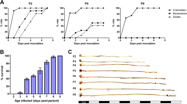
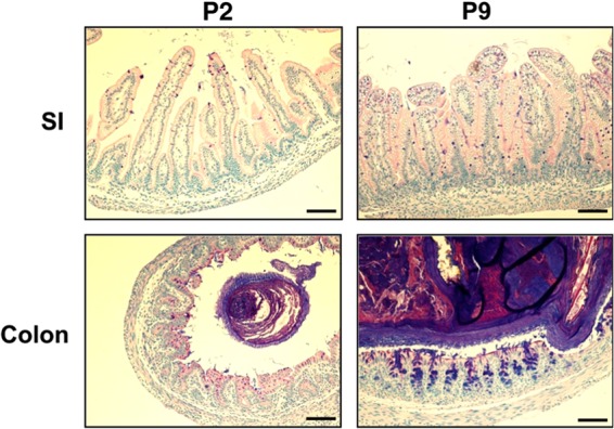
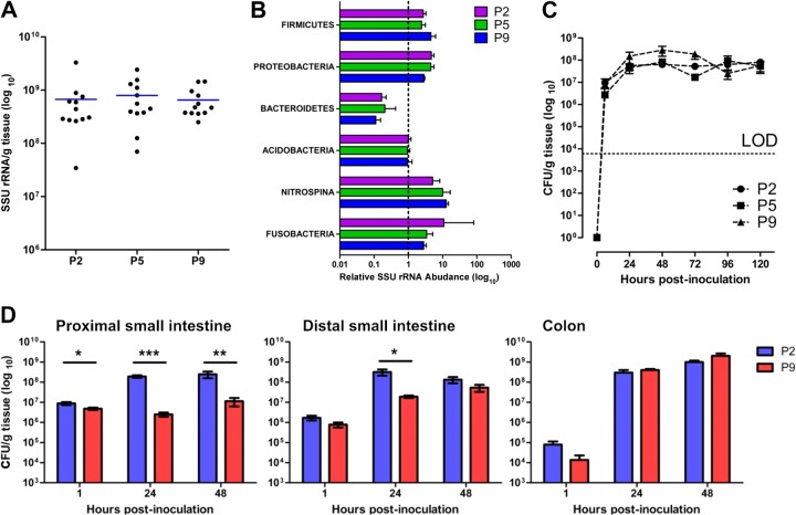
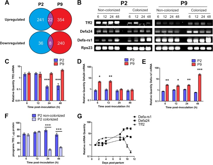
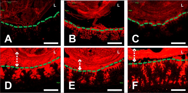
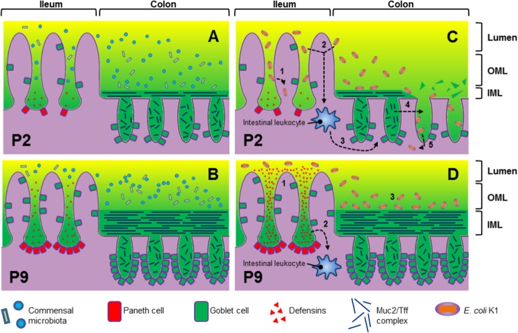
Two-day-old (P2), but not 9-day-old (P9), rat pups are susceptible to systemic infection following gastrointestinal colonization by Escherichia coli K1. Age dependency reflects the capacity of colonizing K1 to translocate from gastrointestinal (GI) tract to blood. A complex GI microbiota developed by P2, showed little variation over P2 to P9, and did not prevent stable K1 colonization. Substantial developmental expression was observed over P2 to P9, including upregulation of genes encoding components of the small intestinal (α-defensins Defa24 and Defa-rs1) and colonic (trefoil factor Tff2) mucus barrier. K1 colonization modulated expression of these peptides: developmental expression of Tff2 was dysregulated in P2 tissues and was accompanied by a decrease in mucin Muc2. Conversely, α-defensin genes were upregulated in P9 tissues. We propose that incomplete development of the mucus barrier during early neonatal life and the capacity of colonizing K1 to interfere with mucus barrier maturation provide opportunities for neuropathogen translocation into the bloodstream.


Figures

Fig 1
Fig 1
Age-dependent susceptibility of the neonatal rat to systemic E. coli K1 infection. (A) GI tract colonization, bacteremia, and accumulated deaths of neonatal rat pups fed E. coli A192PP two (P2), five (P5), and nine (P9) days after birth. All pups in a litter received the oral dose on day 0. Colonies expressing the K1 capsule were detected using K1-specific bacteriophage from perianal swab cultures and from blood samples. The data shown are from representative experiments involving single litters of 9 to 14 pups. (B) Survival of P2-to-P9 rat pups fed E. coli A192PP. Two litters (12 pups per litter) of 2- to 9-day-old pups were used for each time point, and the animals were monitored for a period of 1 week following oral dosing of bacterial cultures; n = 24. Values are means ± standard deviations (SD). (C) Gross morphological appearance of GI tract tissue (stomach to colon) from P2-to-P9 rat pups.
Fig 2
Fig 2
Rapid development of GI tract tissue morphology during the early postnatal period. Small intestinal (SI) and colonic tissues from P2 and P9 animals were fixed, sectioned, and stained with Alcian blue-PAS. The scale bar is 100 μm.
Fig 3
Fig 3
Comparable rates of GI tract colonization by E. coli A192PP over the P2-to-P9 neonatal period complements rapid development of the GI tract microbiota in neonatal rats. (A) Bacterial load, determined by qPCR of conserved 16S SSU rRNA genes, of GI tract tissues and fecal content from P2, P5, and P9 rat pups. At each time point, four animals from each of three litters were sampled. (B) Relative abundance of bacterial phyla of the GI microbiota by SSU rRNA gene microarray. Data were normalized to adult data, as indicated by the dashed line at x = 1. Four litters were employed. Four animals were removed from each litter at P2, P5, and P9; bacterial DNA extracted from tissue samples at each time point hybridized with DNA from the appropriate maternal fecal sample. Results from each of four litters were combined. Probes were ranked according to average Cy5 and Cy3 fluorescence, with the highest shown at the top. (C) Temporal aspects of E. coli K1 GI tract colonization of P2, P5, and P9 animals. K1 bacteria were quantified by qPCR of the neuS gene. DNA was extracted from GI tissues and their contents, and a calibration curve was constructed to convert data to CFU. Two litters of neonatal rats were employed. Four animals were removed from each litter at each time point, the GI tissue was removed and processed, and DNA representing each time point was pooled. LOD, limit of detection. (D) Colonization of the proximal small intestine, distal small intestine, and cecum/colon over the 48-h period following feeding of E. coli A192PP to P2 and P9 pups. E. coli K1 bacteria were quantified by qPCR with the neuS probe. Data were normalized to tissue mass following conversion to CFU. Error bars represent the standard errors of the mean (SEM) from n = 4 animals. Significant differences, as determined by 2-tailed Mann-Whitney test: *, P < 0.05; **, P < 0.01; and ***, P < 0.001.
Fig 4
Fig 4
P2 and P9 host responses to E. coli K1 intestinal colonization. (A) P2 and P9 intestinal tissues show differential transcriptomic responses to E. coli K1 colonization, as demonstrated by comparison of microarray analyses of RNA extracted from colonized and noncolonized P2 and P9 tissues 12 h postinoculation with E. coli K1 or broth. n = 4. (B) Expression of tff2, defa24, defa-rs1, and the reference gene rps23 was analyzed by semiquantitative RT-PCR followed by resolution of pooled cDNA amplicons on agarose gels and by qRT-PCR analysis (C, D, and E) of RNA extracted from GI tract tissues from colonized and noncolonized P2 and P9 animals 6, 12, 24, and 48 h postinoculation with E. coli K1 or broth. Data from colonized animals were normalized to data from an equal number of noncolonized (broth-fed) animals. Tff2 protein was quantified by competitive ELISA of protein extractions from colonized and noncolonized animals (F). The normal developmental expression of tff2, defa24, and defa-rs1 over the period from P1 to P11 was examined by normalizing qRT-PCR data from noncolonized animals to data obtained from RNA extracted from noncolonized P1 neonatal intestinal tissues (G). Error bars for all figures represent the SEM of results from either n = 12 (C, D, and E) or n = 6 (F) animals. Statistically significant differences, as determined by 2-tailed t test, between colonized and noncolonized animals are indicated: *, P < 0.05; **, P < 0.01; ***, P < 0.001.
Fig 5
Fig 5
E. coli K1 colonization of P2 pups results in depletion of Muc2 mucin from colonic epithelial cells. Anti-Muc2 immunostaining of sections of colonic tissues from P2 (A to C) and P9 (D to F) neonatal rats. Tissues were obtained at P2 (A) or P9 (D) and at 48 h after feeding neonates a control dose (B and E) or E. coli A192PP (C and F). The epithelial surface (green dashed lines), lumen (L), and stratified mucus layer (white dashed lines with diamonds) are indicated where appropriate. The scale bars are 100 μm.
Fig 6
Fig 6
Proposed model of GI barrier development in rats from P2 (A) to P9 (B). (C and D) Colonization of the P2 (C) and P9 (D) GI tract by E. coli K1. The numbered steps in panels C and D are expanded in the text.

References

    1. Harvey D, Holt DE, Bedford H. 1999. Bacterial meningitis in the newborn: a prospective study of mortality and morbidity. Semin. Perinatol. 23:218–225 - PubMed
    1. Brouwer MC, Tunkel AR, van de Beek D. 2010. Epidemiology, diagnosis, and antimicrobial treatment of acute bacterial meningitis. Clin. Microbiol. Rev. 23:467–492 - PMC - PubMed
    1. Robbins JB, McCracken GH, Gotschlich EC, Ørskov F, Ørskov I, Hanson LA. 1974. Escherichia coli K1 polysaccharide associated with neonatal meningitis. N. Engl. J. Med. 290:1216–1220 - PubMed
    1. Korhonen TK, Valtonen MV, Parkkinen J, Väisänen-Rhen V, Finne J, Ørskov F, Ørskov I, Svenson SB, Mäkelä PH. 1985. Serotypes, hemolysin production, and receptor recognition of Escherichia coli strains associated with neonatal sepsis and meningitis. Infect. Immun. 48:486–491 - PMC - PubMed
    1. Rutishauser U. 2008. Polysialic acid in the plasticity of the developing and adult vertebrate nervous system. Nat. Rev. Neurosci. 9:26–35 - PubMed

Publication types

MeSH terms