. 2013 Sep;6(5):1167-74.
doi: 10.1242/dmm.011700. Epub 2013 Jun 20.

Dmdmdx/Largemyd: a new mouse model of neuromuscular diseases useful for studying physiopathological mechanisms and testing therapies

Affiliations

Dmdmdx/Largemyd: a new mouse model of neuromuscular diseases useful for studying physiopathological mechanisms and testing therapies

Poliana C M Martins et al. Dis Model Mech. 2013 Sep.

Abstract

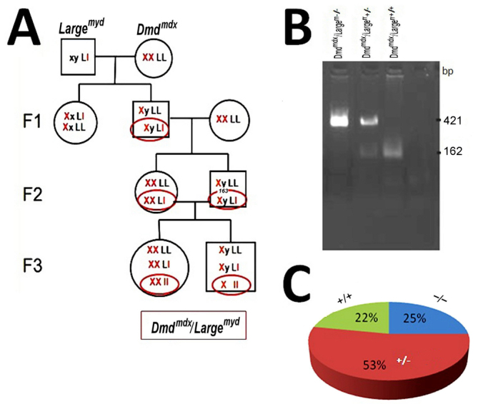
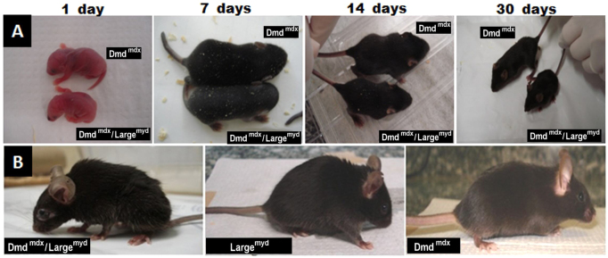
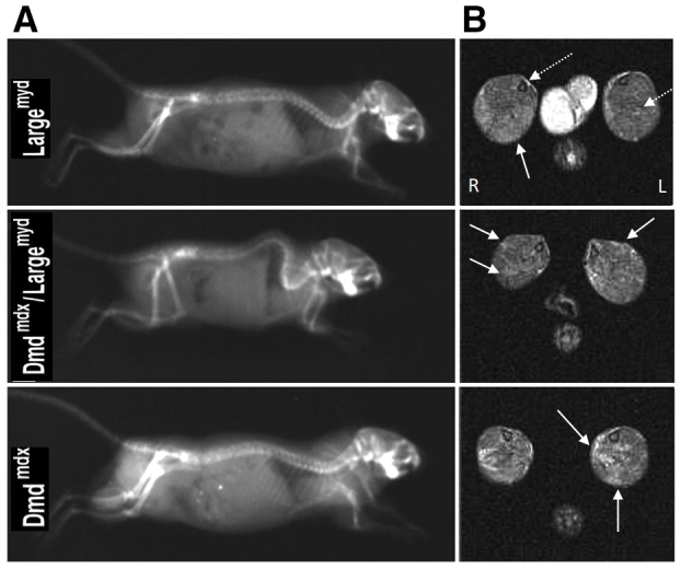
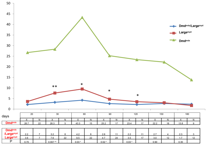
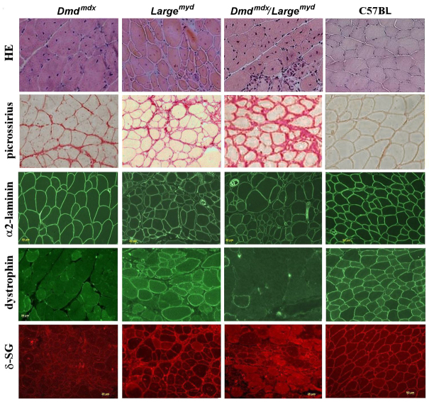
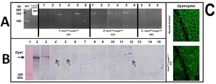
Although muscular dystrophies are among the most common human genetic disorders, there are few treatment options available. Animal models have become increasingly important for testing new therapies prior to entering human clinical trials. The Dmd(mdx) mouse is the most widely used animal model for Duchenne muscular dystrophy (DMD), presenting the same molecular and protein defect as seen in humans with the disease. However, this mouse is not useful for clinical trials because of its very mild phenotype. The mouse model for congenital myodystrophy type 1D, Large(myd), harbors a mutation in the glycosyltransferase Large gene and displays a severe phenotype. To help elucidate the role of the proteins dystrophin and LARGE in the organization of the dystrophin-glycoprotein complex in muscle sarcolemma, we generated double-mutant mice for the dystrophin and LARGE proteins. The new Dmd(mdx)/Large(myd) mouse model is viable and shows a severe phenotype that is associated with the lack of dystrophin in muscle. We tested the usefulness of our new mouse model for cell therapy by systemically injecting them with normal murine mesenchymal adipose stem cells (mASCs). We verified that the mASCs were hosted in the dystrophic muscle. The new mouse model has proven to be very useful for the study of several other therapies, because injected cells can be screened both through DNA and protein analysis. Study of its substantial muscle weakness will also be very informative in the evaluation of functional benefits of these therapies.


Figures

Fig. 1.
Fig. 1.
Generation of the animal model Dmdmdx/Largemyd. (A) The breeding program. L, wild-type Large allele; l, mutated Large allele. (B) The genotyping methodology. The 421 bp band is the mutated allele, and the 162 bp band is the wild-type allele of the Large gene. (C) The proportion of each genotype in F3.
Fig. 2.
Fig. 2.
Phenotypic characterization of the Dmdmdx/Largemyd mouse. (A) Growth and development delay in the Dmdmdx/Largemyd double mutant compared with his brother Dmdmdx at different ages (1, 7, 14 and 30 days). (B) Animals at the age of 120 days.
Fig. 3.
Fig. 3.
Image analyses of the Dmdmdx/Largemyc model. (A) Radiography of the spine of the different models (Largemyd, Dmdmdx/Largemyd and Dmdmdx) at the age of 120 days. (B) Axial T2 weighted MRI of 2-month-old Largemyd, Dmdmdx/Largemyd and Dmdmdx mice, at the lower leg level. Solid arrows indicate affected regions; dashed arrows indicate the position of the lower leg bones, tibia and fibula. L, left side; R, right side.
Fig. 4.
Fig. 4.
Functional analysis of the test bar in Largemyd, Dmdmdx/Largemyd and Dmdmdx mice. x-axis, days; y-axis, the mean time the mice from each group could hold the bar (seconds). The table below the graph shows the mean time the mice from each group could hold the bar (seconds; X), the number of tested animals (N) and the significance (P) in the Mann-Whitney test, comparing the two weaker strains, Dmdmdx/Largemyd versus Largemyd (*P<0.05, **P<0.01).
Fig. 5.
Fig. 5.
H&E staining, picrossirius staining for collagens, and immunohistochemical analysis for α2-laminin, dystrophin and δ-SG in normal C57BL, Dmdmdx, Largemyd and Dmdmdx/Largemyd mice aged 180 days. Magnification 200×.
Fig. 6.
Fig. 6.
DNA screening and protein analyses in the Dmdmdx/Largemyd model. (A) DNA screening for the injected cells: PCR for the Large normal allele (162 bp) in the injected mice. Animals 1 and 2 were injected with cells, and animal 3 with PBS. Columns: 1, heart; 2, gastrocnemius; 3, stomach; 4, quadriceps; 5, diaphragm; 6, tail. Positive control (162 bp) and molecular weight (MW) are shown. (B) Dystrophin western blot analysis from different muscles from the mASC-injected and PBS-injected animals, reacted with antibody to dystrophin (H300, Santa Cruz Biotechnology, Inc.). Column 1, molecular weight; 2, normal human muscle; 3, normal C57BL muscle. Muscles from Dmdmdx/Largemyd mouse 1: column 4, gastrocnemius; 5, quadriceps; 6, diaphragm; 7, heart; 8, stomach. Muscles from Dmdmdx/Largemyd mouse 2: 9, gastrocnemius; 10, quadriceps; 11, diaphragm; 12, heart; 13, stomach. Muscles from Dmdmdx/Largemyd mouse 3: 14, quadriceps; 15, diaphragm. (C) Dystrophin immunofluorescence analysis in a normal control and in one injected muscle: Dmdmdx/Largemyd mouse 2, muscle gastrocnemius.

References

    1. Browning C. A., Grewal P. K., Moore C. J., Hewitt J. E. (2005). A rapid PCR method for genotyping the Large(myd) mouse, a model of glycosylation-deficient congenital muscular dystrophy. Neuromuscul. Disord. 15, 331–335 - PubMed
    1. Bulfield G., Siller W. G., Wight P. A., Moore K. J. (1984). X chromosome-linked muscular dystrophy (mdx) in the mouse. Proc. Natl. Acad. Sci. USA 81, 1189–1192 - PMC - PubMed
    1. Campbell K. P., Kahl S. D. (1989). Association of dystrophin and an integral membrane glycoprotein. Nature 338, 259–262 - PubMed
    1. Cohn R. D., Campbell K. P. (2000). Molecular basis of muscular dystrophies. Muscle Nerve 23, 1456–1471 - PubMed
    1. Dangain J., Vrbova G. (1984). Muscle development in mdx mutant mice. Muscle Nerve 7, 700–704 - PubMed

Publication types

MeSH terms