. 2013 Aug 2;288(31):22516-26.
doi: 10.1074/jbc.M113.489328. Epub 2013 Jun 24.

Inhibition of HDAC6 deacetylase activity increases its binding with microtubules and suppresses microtubule dynamic instability in MCF-7 cells

Affiliations

Inhibition of HDAC6 deacetylase activity increases its binding with microtubules and suppresses microtubule dynamic instability in MCF-7 cells

Jayant Asthana et al. J Biol Chem. .

Abstract

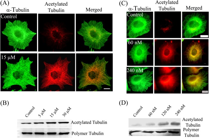
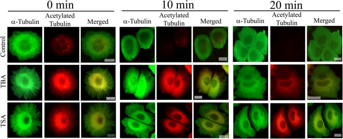
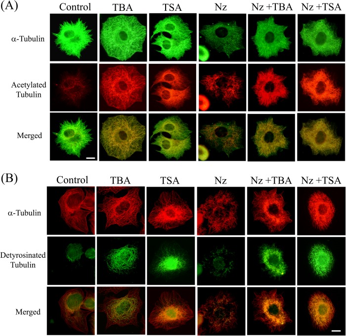
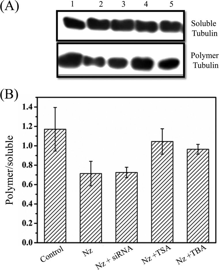
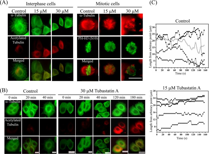
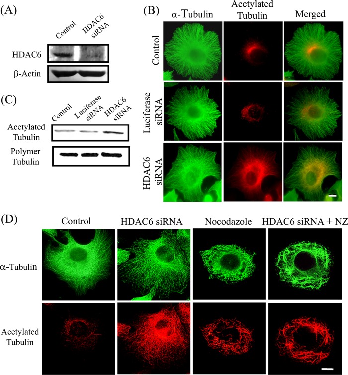
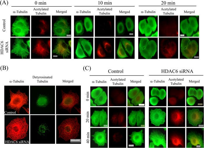
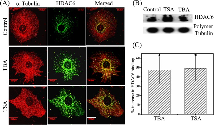
The post-translational modification of tubulin appears to be a highly controlled mechanism that regulates microtubule functioning. Acetylation of the ε-amino group of Lys-40 of α-tubulin marks stable microtubules, although the causal relationship between tubulin acetylation and microtubule stability has remained poorly understood. HDAC6, the tubulin deacetylase, plays a key role in maintaining typical distribution of acetylated microtubules in cells. Here, by using tubastatin A, an HDAC6-specific inhibitor, and siRNA-mediated depletion of HDAC6, we have explored whether tubulin acetylation has a role in regulating microtubule stability. We found that whereas both pharmacological inhibition of HDAC6 as well as its depletion enhance microtubule acetylation, only pharmacological inhibition of HDAC6 activity leads to an increase in microtubule stability against cold and nocodazole-induced depolymerizing conditions. Tubastatin A treatment suppressed the dynamics of individual microtubules in MCF-7 cells and delayed the reassembly of depolymerized microtubules. Interestingly, both the localization of HDAC6 on microtubules and the amount of HDAC6 associated with polymeric fraction of tubulin were found to increase in the tubastatin A-treated cells compared with the control cells, suggesting that the pharmacological inhibition of HDAC6 enhances the binding of HDAC6 to microtubules. The evidence presented in this study indicated that the increased binding of HDAC6, rather than the acetylation per se, causes microtubule stability. The results are in support of a hypothesis that in addition to its deacetylase function, HDAC6 might function as a MAP that regulates microtubule dynamics under certain conditions.

Keywords: Histone Deacetylase; Histone Deacetylase 6; Histone Deacetylase Inhibitors; MAPs; Microtubule Dynamics; Microtubules; Post-translational Modification; Tubastatin A; Tubulin Acetylation.


Figures

FIGURE 1.
FIGURE 1.
Tubastatin A and TSA increased the microtubule acetylation level. Effects of tubastatin A (A and B) and TSA (C and D) on microtubules and microtubule acetylation level are shown. A and C, MCF-7 cells were treated with different concentrations of tubastatin A (A) or of TSA (C) for 24 h and processed for immunostaining with antibodies against α-tubulin (green) and acetylated tubulin (red). Scale bars, 10 μm. B and D, MCF-7 cells were treated with different concentrations of tubastatin A (B) or of TSA (D) for 24 h. Polymeric fractions of tubulin were isolated, processed for Western blotting, and were probed for α-tubulin and acetylated tubulin.
FIGURE 2.
FIGURE 2.
Tubastatin A and TSA stabilized microtubules against cold-induced depolymerization. MCF-7 cells were treated with vehicle, 30 μm tubastatin A (TBA), or 240 nm TSA for 24 h. Then cells were incubated on ice to depolymerize microtubules and were fixed at the indicated time points (0, 10, and 20 min). Fixed cells were processed for immunostaining with antibodies against α-tubulin (green) and acetylated tubulin (red). Scale bars, 10 μm.
FIGURE 3.
FIGURE 3.
Tubastatin A and TSA stabilized microtubules against nocodazole-induced disassembly. A, MCF-7 cells were treated with either vehicle, 15 μm tubastatin A (TBA), 240 nm TSA, or 200 nm nocodazole (Nz) individually or with a combination of nocodazole (200 nm) and tubastatin A (15 μm) or nocodazole (200 nm) and TSA (240 nm) for 24 h and processed for immunostaining with antibodies against α-tubulin (green) and acetylated tubulin (red). Scale bar, 10 μm. B, MCF-7 cells were treated with either vehicle, 15 μm tubastatin A (TBA), 240 nm TSA, 200 nm nocodazole (Nz) individually or with a combination of nocodazole (200 nm) and tubastatin A (15 μm) or nocodazole (200 nm) and TSA (240 nm) for 24 h and immunostained with antibodies against α-tubulin (red) and detyrosinated tubulin (green). Scale bar, 10 μm.
FIGURE 4.
FIGURE 4.
Effects of tubastatin A, TSA, and HDAC6 siRNA on nocodazole-induced depolymerization of microtubules. A, polymeric and soluble tubulin fractions of MCF-7 cells treated with vehicle (lane 1), 200 nm nocodazole (Nz) alone (lane 2), or 200 nm nocodazole in combination with HDAC6 siRNA, 240 nm TSA, or 15 μm tubastatin A (TBA) (lanes 3, 4 and 5, respectively) for 24 h were isolated as described, and equal amounts of proteins were resolved by SDS-PAGE followed by immunoblotting with anti-α-tubulin antibody. B, the ratio of polymer to soluble fraction of tubulin in cells treated as in A was measured from the intensity of the bands in blot. Data were an average of three independent experiments and represent mean ± S.D. (error bars).
FIGURE 5.
FIGURE 5.
Tubastatin A altered the assembly dynamics of interphase microtubules in MCF-7 cells. A, tubastatin A delayed the reassembly of nocodazole-depolymerized interphase and mitotic microtubules. Cells were treated with either 500 nm nocodazole for 4 h (interphase cells) or 300 nm nocodazole for 24 h (mitotic cells). Nocodazole was then removed by repeated washing, and microtubules were allowed to reassemble in the absence (control) or presence of 15 or 30 μm tubastatin A for 90 min. Cells were then fixed and processed for co-immunostaining with antibodies against α-tubulin (green) and acetylated tubulin (red) (interphase cells) or α-tubulin (red) and phosphohistone-H3 (Ser-10) (green) (mitotic cells). PH-H3 (Ser-10), a mitotic marker, was used to indicate the chromosomes. Scale bar, 10 μm. B, tubastatin A delayed the reassembly of cold-depolymerized interphase microtubules. Microtubules were depolymerized by incubating the cells on ice for 45 min. Then cells were incubated with prewarmed medium, without or with 30 μm tubastatin A, at 37 °C in incubator for different time intervals (0, 20, 40, 120, and 180 min) and then fixed and processed for immunostaining with antibodies against α-tubulin (green) and acetylated tubulin (red). Scale bars, 10 μm. C, life history plots of the individual microtubules in EGFP-tubulin-expressing MCF-7 cells treated with vehicle or 15 μm tubastatin A for 24 h. The initial length represents length from an arbitrary fixed point of microtubule to the end of the microtubule.
FIGURE 6.
FIGURE 6.
Effect of depletion of HDAC6 on microtubule stability. A, levels of HDAC6 in cells treated with luciferase siRNA (control) and siRNA against HDAC6. β-Actin was used as loading control. B, effect of depletion of HDAC6 on microtubules and acetylated tubulin. MCF-7 cells were kept untreated or were treated with siRNA against luciferase or HDAC6 and were then processed for immunostaining with antibodies against α-tubulin (green) and acetylated tubulin (red). Scale bar, 10 μm. C, MCF-7 cells untreated or treated with siRNA against luciferase or HDAC6. The polymeric fractions of tubulin were isolated from the cells. The samples were processed for Western blotting and were probed for α-tubulin and acetylated tubulin. D, effect of depletion of HDAC6 on nocodazole-induced microtubule depolymerization. MCF-7 cells were treated with HDAC6 siRNA and 200 nm nocodazole alone or together with HDAC6 siRNA for 24 h. The cells were then fixed and processed for immunostaining with antibodies against α-tubulin (green) and acetylated tubulin (red). Scale bar, 10 μm.
FIGURE 7.
FIGURE 7.
Depletion of HDAC6 did not stabilize microtubules. A, effect of depletion of HDAC6 on stability of microtubules against cold-induced depolymerization. MCF-7 cells were transfected with luciferase siRNA (control) or HDAC6 siRNA for 24 h. Then cells were incubated on ice to depolymerize microtubules and were fixed at the indicated time points (0, 10, and 20 min). Fixed cells were processed for immunostaining with antibodies against α-tubulin (green) and acetylated tubulin (red). Scale bars, 10 μm. B, MCF-7 cells treated with luciferase siRNA (control) or HDAC6 siRNA for 24 h. After treatment, cells were fixed and processed for immunostaining with antibodies against α-tubulin (red) and detyrosinated tubulin (green). Scale bar, 10 μm. C, depletion of HDAC6 not delaying reassembly of cold-depolymerized interphase microtubules. MCF-7 cells were treated with luciferase siRNA (control) or HDAC6 siRNA for 24 h. After treatment, microtubules were depolymerized by incubating the cells on ice for 45 min. Then cells were incubated with prewarmed medium at 37 °C in an incubator for different time intervals and then fixed and processed for immunostaining with antibodies against α-tubulin (green) and acetylated tubulin (red). The status of microtubules and their acetylation level in cells at 0, 20, and 40 min is shown. Scale bars, 10 μm.
FIGURE 8.
FIGURE 8.
Tubastatin A and TSA increased the binding of HDAC6 with interphase microtubules in MCF-7 cells. A, cells treated with vehicle, 30 μm tubastatin A (TBA), or 240 nm TSA for 24 h and fixed and processed to visualize microtubules (red) and HDAC6 (green). Scale bar, 20 μm. B, Western blot analysis of HDAC6 bound with the polymeric fraction of tubulin in cells. MCF-7 cells were treated with vehicle, 240 nm TSA, or 30 μm tubastatin A for 24 h. The polymeric fraction of tubulin was isolated and probed with antibodies against α-tubulin and HDAC6. C, histogram showing the percentage increase in the binding of HDAC6 with polymeric tubulin after treatment of cells with tubastatin A and TSA with respect to control as quantified from B. Data are average of five independent set of experiments and represent mean ± S.D. (error bars). *, p ≤ 0.001.

References

    1. Janke C., Bulinski J. C. (2011) Post-translational regulation of the microtubule cytoskeleton: mechanisms and functions. Nat. Rev. Mol. Cell Biol. 12, 773–786 - PubMed
    1. Akella J. S., Wloga D., Kim J., Starostina N. G., Lyons-Abbott S., Morrissette N. S., Dougan S. T., Kipreos E. T., Gaertig J. (2010) MEC-17 is an α-tubulin acetyltransferase. Nature 467, 218–222 - PMC - PubMed
    1. Hubbert C., Guardiola A., Shao R., Kawaguchi Y., Ito A., Nixon A., Yoshida M., Wang X. F., Yao T. P. (2002) HDAC6 is a microtubule-associated deacetylase. Nature 417, 455–458 - PubMed
    1. Matsuyama A., Shimazu T., Sumida Y., Saito A., Yoshimatsu Y., Seigneurin-Berny D., Osada H., Komatsu Y., Nishino N., Khochbin S., Horinouchi S., Yoshida M. (2002) In vivo destabilization of dynamic microtubules by HDAC6-mediated deacetylation. EMBO J. 21, 6820–6831 - PMC - PubMed
    1. North B. J., Marshall B. L., Borra M. T., Denu J. M., Verdin E. (2003) The human Sir2 ortholog, SIRT2, is an NAD+-dependent tubulin deacetylase. Mol. Cell 11, 437–444 - PubMed

Publication types