Disseminated histoplasmosis with oral and cutaneous manifestations
- PMID: 23798850
- PMCID: PMC3687172
- DOI: 10.4103/0973-029X.110722
Disseminated histoplasmosis with oral and cutaneous manifestations
Abstract
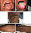
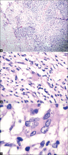
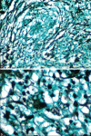
Histoplasmosis is a systemic mycotic infection caused by the dimorphic fungus, Histoplasma capsulatum. Systemic histoplasmosis has emerged as an important opportunistic infection in human immunodeficiency virus (HIV) patients and those in endemic areas. Reported cases of histoplasmosis have been low in India with less than 50 cases being reported. We are reporting a case of disseminated histoplasmosis with oral and cutaneous involvement in an HIV seronegative patient.
Keywords: Disseminated; histoplasmosis; human immunodeficiency virus.
Conflict of interest statement
Figures


References
-
- Mignogna MD, Fedele S, Lo Russo L, Ruoppo E, Lo Muzio L. A case of oral localized histoplasmosis in an immunocompetent patient. Eur J Clin Microbiol Infect Dis. 2001;20:753–5. - PubMed
-
- Rappo U, Beitler JR, Faulhaber JR, Firoz B, Henning JS, Thomas KM, et al. Expanding the horizons of histoplasmosis: Disseminated histoplasmosis in a renal transplant patient after a trip to Bangladesh. Transpl Infect Dis. 2010;12:155–60. - PubMed
-
- Ferreira OG, Cardoso SV, Borges AS, Ferreira MS, Loyola AM. Oral histoplasmosis in Brazil. Oral Surg Oral Med Oral Pathol Oral Radiol Endod. 2002;93:654–9. - PubMed
-
- Hernández SL, López De Blanc SA, Sambuelli RH, Roland H, Cornelli C, Lattanzi V, et al. Oral histoplasmosis associated with HIV infection: A comparative study. J Oral Pathol Med. 2004;33:445–50. - PubMed
Publication types
LinkOut - more resources
Full Text Sources
Other Literature Sources

