. 2013 Jun 14;8(6):e65549.
doi: 10.1371/journal.pone.0065549. Print 2013.

Adaptive induction of growth differentiation factor 15 attenuates endothelial cell apoptosis in response to high glucose stimulus

Affiliations

Adaptive induction of growth differentiation factor 15 attenuates endothelial cell apoptosis in response to high glucose stimulus

Jun Li et al. PLoS One. .

Abstract

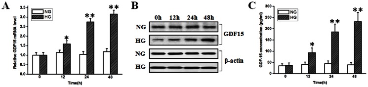
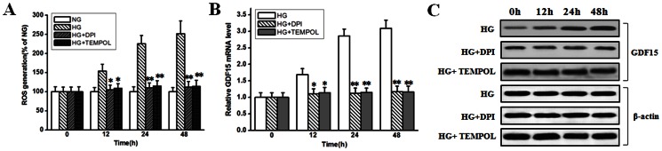
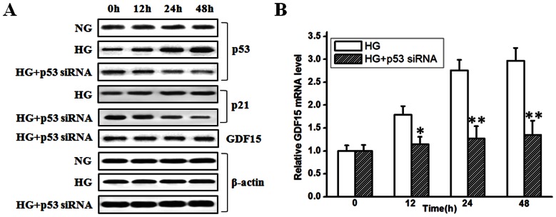
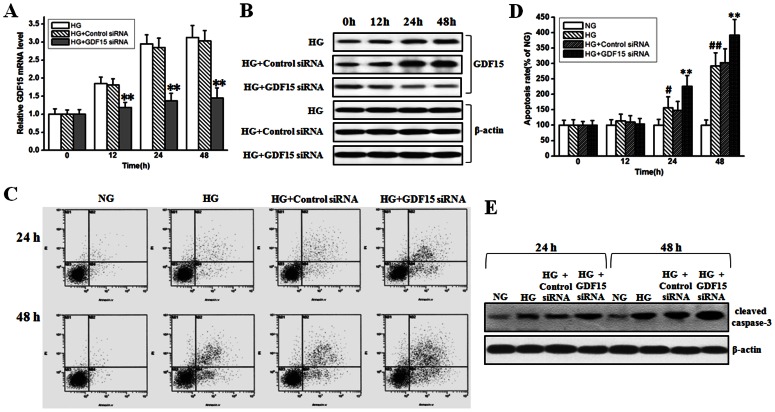
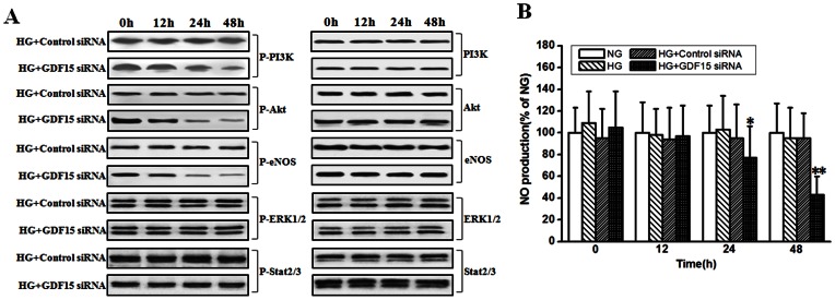
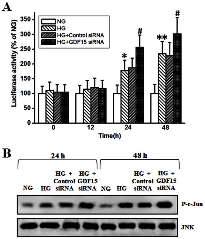
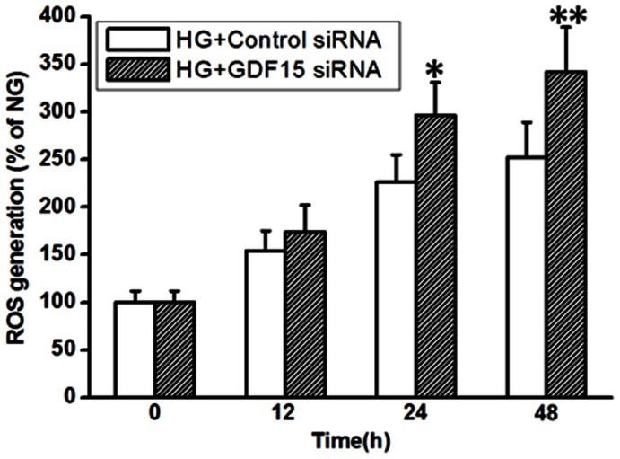
Growth differentiation factor 15 (GDF15), a direct target gene of p53, is a multifunctional member of the TGF-β/BMP superfamily. GDF15 can be induced and is implicated as a key secretory cytokine in response to multiple cellular stimuli. Accumulating evidence indicates that GDF15 is associated with the development and prognosis of diabetes mellitus, while whether GDF15 can be induced by high glucose is unknown. In the present study, we revealed that high glucose could induce GDF15 expression and secretion in cultured human umbilical vein endothelial cells in a ROS- and p53-dependent manner. Inhibition of high glucose-induced GDF15 expression by siRNA demonstrated that adaptively induced GDF15 played a protective role against high glucose-induced human umbilical vein endothelial cell apoptosis via maintaining the active state of PI3K/Akt/eNOS pathway and attenuating NF-κB/JNK pathway activation. The protective effects of GDF15 were probably achieved by inhibiting ROS overproduction in high glucose-treated human umbilical vein endothelial cells in a negative feedback manner. Our results suggest that high glucose can promote GDF15 expression and secretion in human umbilical vein endothelial cells, which in turn attenuates high glucose-induced endothelial cell apoptosis.


Conflict of interest statement

Competing Interests: The authors have declared that no competing interests exist.

Figures

Figure 1
Figure 1. Adaptive induction of GDF15 in HUVEC cells in response to high glucose stimulus.
HUVEC cells were treated with normal (5.5 mmol/l) or high (33.3 mmol/l) glucose. At indicated time points, GDF15 expression and secretion were detected by RT-PCR, western blot, and ELISA. A, analysis of GDF15 mRNA expression with RT-PCR. B, western blots of GDF15 and β-actin. C, ELISA assay of secreted GDF15. NG, normal glucose. HG, high glucose. *, P<0.05, **, P<0.01 versus NG.
Figure 2
Figure 2. ROS inhibitors abolished high glucose-induced GDF15 expression.
HUVEC cells were treated with normal (5.5 mmol/l) or high (33.3 mmol/l) glucose in the absence or presence of 10 µmol/l DPI or 1 mmol/l TEMPOL. A, ROS production were assayed as described in the materials and methods. B, C, at indicated time points, GDF15 mRNA and protein expression were detected by RT-PCR and western blot. NG, normal glucose. HG, high glucose. *, P<0.05, **, P<0.01 versus HG.
Figure 3
Figure 3. Knockdown of p53 expression inhibited high glucose-induced GDF15 expression.
p53 siRNA was transfected into HUVEC cells. 12 h after transfection, HUVEC cells were treated with high glucose (33.3 mmol/l). A, at indicated time points, p53, p21, GDF15, and β-actin proteins were detected by western blot. B, GDF15 mRNA was detected by RT-PCR. NG, normal glucose. HG, high glucose. *, P<0.05, **, P<0.01 versus HG.
Figure 4
Figure 4. Inhibition of adaptively induced GDF15 expression enhanced high glucose-induced HUVEC cell apoptosis.
GDF15 siRNA or negative control siRNA were transfected into HUVEC cells. 12 h after transfection, HUVEC cells were treated with normal (5.5 mmol/l) or high (33.3 mmol/l) glucose. A, B, mRNA and protein levels of GDF15 was detected to test the efficiency of GDF15 siRNA. C, D, FCM results and statistical analysis of high glucose-induced HUVEC cell apoptosis with or without GDF15 siRNA transfection. E, high glucose-induced caspase-3 cleavage with or without GDF15 siRNA transfection. NG, normal glucose. HG, high glucose. *, P<0.05, **, P<0.01 versus HG. #, P<0.05, ##, P<0.01 versus NG.
Figure 5
Figure 5. Inhibition of adaptively induced GDF15 expression attenuated PI3K/Akt/eNOS pathway.
GDF15 siRNA or negative control siRNA were transfected into HUVEC cells. 12 h after transfection, HUVEC cells were treated with high (33.3 mmol/l) glucose. A, at indicated time points, phosphorylation and total amount of PI3K, Akt, eNOS, ERK1/2, and Stat2/3 were detected by western blot. B, The concentrations of NO in the culture medium were measured using the nitric oxide colorimetric assay kit as described in the materials and methods. NG, normal glucose. HG, high glucose. *, P<0.05, **, P<0.01 versus HG.
Figure 6
Figure 6. Inhibition of adaptively induced GDF15 expression promoted NF-κB/JNK activation.
GDF15 siRNA or negative control siRNA were transfected into HUVEC cells. 12 h after transfection, HUVEC cells were treated with normal (5.5 mmol/l) or high (33.3 mmol/l) glucose. A, at indicated time points, NF-κB luciferase activity was detected using a Cignal NF-κB reporter (luc) kit as described in the materials and methods. B, JNK activity was detected by a JNK activity assay kit using recombinant c-Jun as substrate as described in the materials and methods. NG, normal glucose. HG, high glucose. *, P<0.05, **, P<0.01 versus NG. #, P<0.05 versus HG.
Figure 7
Figure 7. Inhibition of adaptively induced GDF15 expression increased ROS production.
GDF15 siRNA or negative control siRNA were transfected into HUVEC cells. 12 h after transfection, HUVEC cells were treated with high (33.3 mmol/l) glucose. At indicated time points, ROS production were assayed as described in the materials and methods. HG, high glucose. *, P<0.05, **, P<0.01 versus Control siRNA.

References

    1. Bootcov MR, Bauskin AR, Valenzuela SM, Moore AG, Bansal M, et al. (1997) MIC-1, a novel macrophage inhibitory cytokine, is a divergent member of the TGF-beta superfamily. Proc Natl Acad Sci U S A 94: 11514–11519. - PMC - PubMed
    1. Husaini Y, Qiu MR, Lockwood GP, Luo XW, Shang P, et al. (2012) Macrophage inhibitory cytokine-1 (MIC-1/GDF15) slows cancer development but increases metastases in TRAMP prostate cancer prone mice. PLoS One 7: e43833. - PMC - PubMed
    1. Lawton LN, Bonaldo MF, Jelenc PC, Qiu L, Baumes SA, et al. (1997) Identification of a novel member of the TGF-beta superfamily highly expressed in human placenta. Gene 203: 17–26. - PubMed
    1. Yokoyama-Kobayashi M, Saeki M, Sekine S, Kato S (1997) Human cDNA encoding a novel TGF-beta superfamily protein highly expressed in placenta. J Biochem 122: 622–626. - PubMed
    1. Tan M, Wang Y, Guan K, Sun Y (2000) PTGF-beta, a type beta transforming growth factor (TGF-beta) superfamily member, is a p53 target gene that inhibits tumor cell growth via TGF-beta signaling pathway. Proc Natl Acad Sci U S A 97: 109–114. - PMC - PubMed

Publication types

MeSH terms