. 2013 Jun 14;8(6):e65809.
doi: 10.1371/journal.pone.0065809. Print 2013.

Distinctive profile of IsomiR expression and novel microRNAs in rat heart left ventricle

Affiliations

Distinctive profile of IsomiR expression and novel microRNAs in rat heart left ventricle

Mary K McGahon et al. PLoS One. .

Abstract

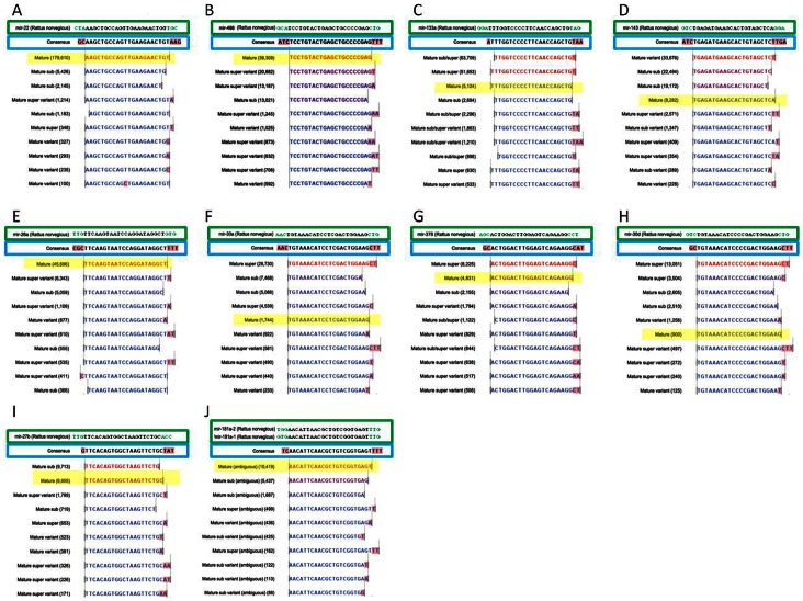
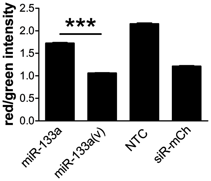
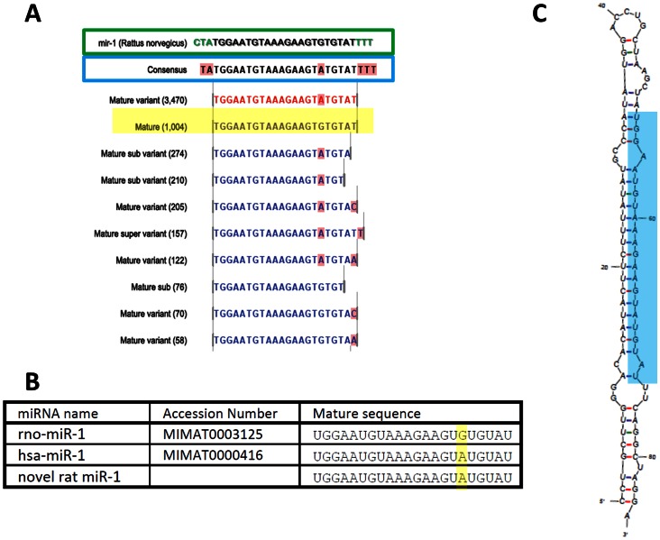
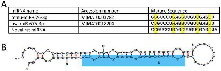
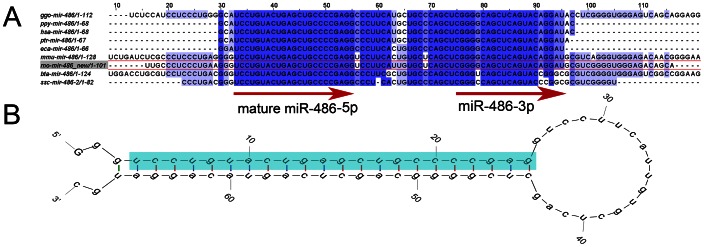
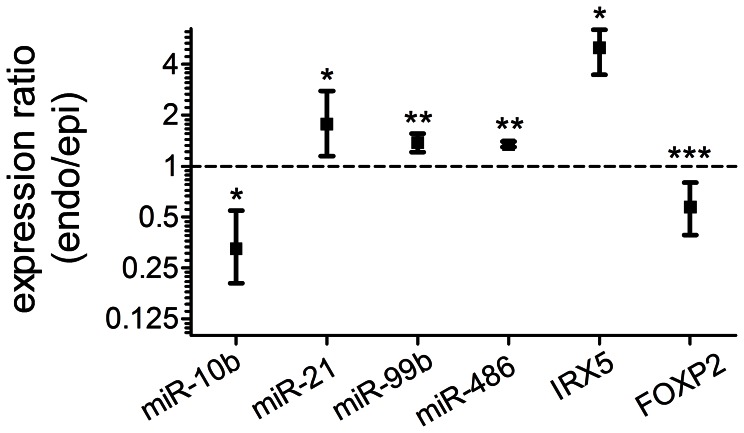
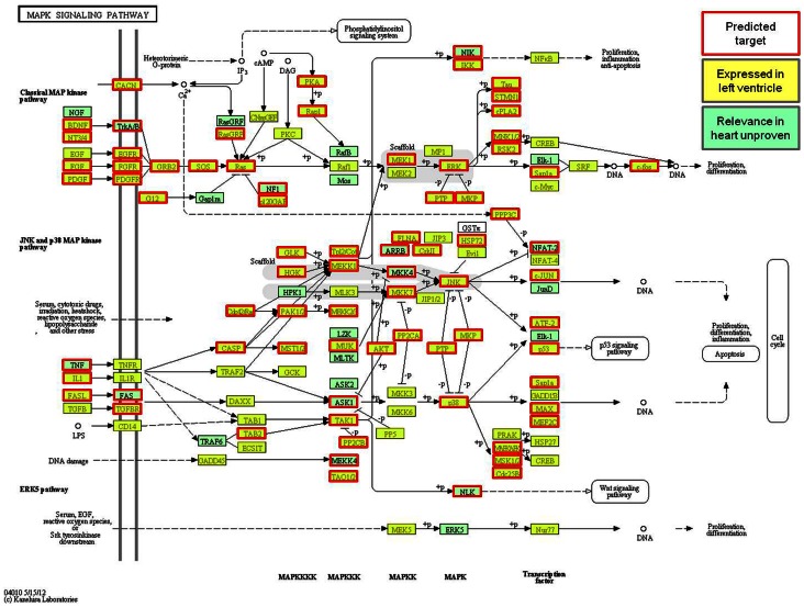
MicroRNAs (miRNAs) are single-stranded non-coding RNAs that negatively regulate target gene expression through mRNA cleavage or translational repression. There is mounting evidence that they play critical roles in heart disease. The expression of known miRNAs in the heart has been studied at length by microarray and quantitative PCR but it is becoming evident that microRNA isoforms (isomiRs) are potentially physiologically important. It is well known that left ventricular (patho)physiology is influenced by transmural heterogeneity of cardiomyocyte phenotype, and this likely reflects underlying heterogeneity of gene expression. Given the significant role of miRNAs in regulating gene expression, knowledge of how the miRNA profile varies across the ventricular wall will be crucial to better understand the mechanisms governing transmural physiological heterogeneity. To determinine miRNA/isomiR expression profiles in the rat heart we investigated tissue from different locations across the left ventricular wall using deep sequencing. We detected significant quantities of 145 known rat miRNAs and 68 potential novel orthologs of known miRNAs, in mature, mature* and isomiR formation. Many isomiRs were detected at a higher frequency than their canonical sequence in miRBase and have different predicted targets. The most common miR-133a isomiR was more effective at targeting a construct containing a sequence from the gelsolin gene than was canonical miR-133a, as determined by dual-fluorescence assay. We identified a novel rat miR-1 homolog from a second miR-1 gene; and a novel rat miRNA similar to miR-676. We also cloned and sequenced the rat miR-486 gene which is not in miRBase (v18). Signalling pathways predicted to be targeted by the most highly detected miRNAs include Ubiquitin-mediated Proteolysis, Mitogen-Activated Protein Kinase, Regulation of Actin Cytoskeleton, Wnt signalling, Calcium Signalling, Gap junctions and Arrhythmogenic Right Ventricular Cardiomyopathy. Most miRNAs are not expressed in a gradient across the ventricular wall, with exceptions including miR-10b, miR-21, miR-99b and miR-486.


Conflict of interest statement

Competing Interests: The authors have declared that no competing interests exist.

Figures

Figure 1
Figure 1. Length distribution analysis of small RNA sequences in 3 mid-myocardial rat samples.
Reads greater than 12 nt are included. Red bars represent reads annotated to known rat miRNAs (miRBase v18), green bars represent reads annotated to miRNAs in other species (novel orthologs) and black bars represent unannotated small RNA sequences.
Figure 2
Figure 2. miRNA detection frequency.
Most highly detected miRNAs grouped on mature sequence, normalised to total annotated reads (expressed in reads per million mapped (RPMM) from 3 rat mid-myocardial samples).
Figure 3
Figure 3. Frequency of isomiRs with 5′ and/or 3′ variations.
Distributions of 5′ (left) and 3′ (right) end variants are shown for miRNAs derived from the 5′ arm (A) and from the 3′ arm (B) of the pre-miR hairpin. Data are mean ± SEM for three mid-myocardial samples.
Figure 4
Figure 4. IsomiRs of selected miRNAs.
IsomiR sequences of the 10 most highly detected miRNAs (mature miRNA highlighted in yellow) with expression values in brackets from a mid-myocardial sample aligned to the published pre-miR sequence (boxed in green; miRBase v18). Consensus sequence (boxed in blue) for each miRNA represents the most prevalent nt aligned at each position (nts highlighted in pink represent variations from the miRBase published mature sequence).
Figure 5
Figure 5. Differential suppression of a gelsolin sequence-tagged reporter gene by miR-133a isomiRs.
HEK293 cells were transfected with expression plasmids encoding mCherry with a 3′ partial gelsolin sequence and pSM30 containing inserts for miR-133a, miR-133a(v), a random non-targeting sequence (NTC) or siRNA against mCherry (siR-mCh). Cells were imaged and analysed for both green and red fluorescence intensity. A decrease in red/green ratio vs NTC indicates downregulation of target gene expression. Data were log transformed and compared by one-way ANOVA with Bonferroni's Multiple Comparison Test. All pairwise comparisons were significant (p<.001). Comparison of miR-133a vs miR-133a(v) is indicated (***). Number of cells (n): miR-133a 4640; miR-133a(v) 2843; NTC 4088; siR-mCh 3884. Representative of 3 separate experiments in which miR-133a(v) was significantly more effective than miR-133a.
Figure 6
Figure 6. Relative miRNA abundance by TaqMan assay.
Expression of miRNAs relative to rno-miR-1 according to 2−ΔCt where ΔCt = Ct – CtmiR-1 (mean ± SEM). Ct values were compared by repeated measures ANOVA and Tukey’s multiple comparison test; **p<.01, ***p<.001. Mid-myocardial samples from n = 6 hearts.
Figure 7
Figure 7. Novel rno-miR-1 sequence as detected by deep sequencing.
A: Ten most frequently detected isomiR sequences of miR-1 (rat mature miRNA highlighted in yellow) with expression values in brackets aligned to the published pre-miR sequence (boxed in green; miRBase v18). The consensus sequence (boxed in blue) represents the most prevalent nt aligned at each position (nts highlighted in pink represent variations from the miRBase published mature sequence). B: miR-1 sequences as published in miRBase v18 showing the novel rat miR-1 sequence aligned with the previously reported rat miR-1 and the human sequence (with which it is identical). C: Predicted stem-loop structure (mFold 3.2) of proposed pre-miR deduced from genomic sequence. Mature product highlighted in blue.
Figure 8
Figure 8. Novel rat miRNA similar to miR-676-3p.
A: Alignment of the novel rat miRNA sequence to miR-676-3p sequences as published in miRBase v18. B: Predicted stem-loop structure (mFold 3.2) of proposed pre-miR deduced from genomic sequence. Mature miRNA highlighted in blue.
Figure 9
Figure 9. The rat miR-486 gene.
A: Alignment of the genomic miR-486 sequence across nine species including rat (highlighted in red). Blue shading indicates percentage sequence identity and the mature and ‘star’ sequences represented by the most common reads are indicated with arrows. B: Predicted stem-loop structure (mFold 3.2) of pre-miR-486 deduced from genomic sequence. Mature miRNA highlighted in blue.
Figure 10
Figure 10. Transmural miRNA expression gradients.
Taqman small RNA assays and TaqMan gene expression assays were performed on epicardial and endocardial samples from rat hearts. Endocardial/epicardial expression ratios were determined by analysis of Ct values using REST 2009. The reference genes utilised were miRNAs with stable expression across samples according to BestKeeper: miR-22, miR-30a, miR-30c, miR-30e and miR-100. The reference genes for IRX5 and FOXP2 were GAPDH, HPRT1 and 18s rRNA. Data are mean ± SEM; n = 4 hearts for miRNAs (n = 3 of these for IRX5 and FOXP2). *P<.05; **P<.01; ***P<.001.
Figure 11
Figure 11. Pathway analysis of MAP Kinase signaling (Kegg pathway).
Highlighted genes are targets predicted by 2 or more algorithms of the top 16 highest detected miRNAs (boxed in red; P = 1.86×10⁻⁶; 75 targets) showing overlap with genes expressed in the heart (highlighted; expression data 9 SD rat left ventricular myocardium samples from 2 microarray experiments, (GSE6943; GSM160095–100 & GSE6880; GSM158589–91; http://www.ncbi.nlm.nih.gov/gds).

References

    1. Bartel DP (2004) MicroRNAs: genomics, biogenesis, mechanism, and function. Cell 116: 281–297. - PubMed
    1. D'Alessandra Y, Pompilio G, Capogrossi MC (2012) MicroRNAs and myocardial infarction. Curr Opin Cardiol 27: 228–235. - PubMed
    1. Bostjancic E, Zidar N, Stajer D, Glavac D (2010) MicroRNAs miR-1, miR-133a, miR-133b and miR-208 are dysregulated in human myocardial infarction. Cardiology 115: 163–169. - PubMed
    1. Gladka MM, da Costa Martins PA, De Windt LJ (2012) Small changes can make a big difference - microRNA regulation of cardiac hypertrophy. J Mol Cell Cardiol 52: 74–82. - PubMed
    1. D'Alessandra Y, Devanna P, Limana F, Straino S, Di Carlo A, et al. (2010) Circulating microRNAs are new and sensitive biomarkers of myocardial infarction. Eur Heart J 31: 2765–2773. - PMC - PubMed

Publication types

MeSH terms