

Evaluating amyloid-β oligomers in cerebrospinal fluid as a biomarker for Alzheimer's disease
- PMID: 23799095
- PMCID: PMC3682966
- DOI: 10.1371/journal.pone.0066381
Evaluating amyloid-β oligomers in cerebrospinal fluid as a biomarker for Alzheimer's disease
Abstract
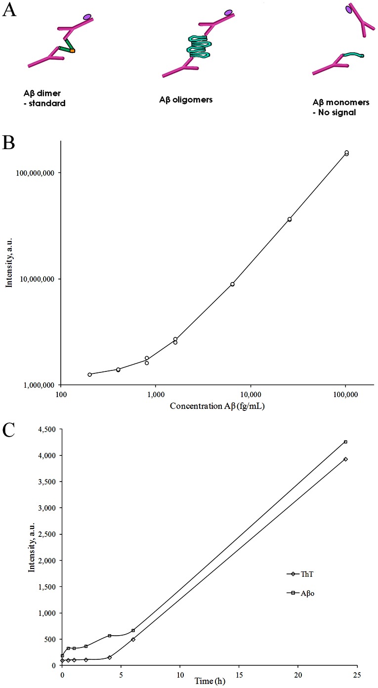
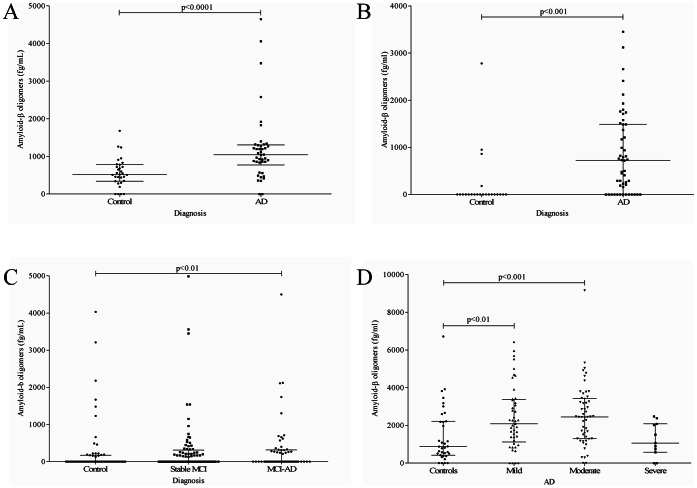
The current study evaluated amyloid-β oligomers (Aβo) in cerebrospinal fluid as a clinical biomarker for Alzheimer's disease (AD). We developed a highly sensitive Aβo ELISA using the same N-terminal monoclonal antibody (82E1) for capture and detection. CSF samples from patients with AD, mild cognitive impairment (MCI), and healthy controls were examined. The assay was specific for oligomerized Aβ with a lower limit of quantification of 200 fg/ml, and the assay signal showed a tight correlation with synthetic Aβo levels. Three clinical materials of well characterized AD patients (n = 199) and cognitively healthy controls (n = 148) from different clinical centers were included, together with a clinical material of patients with MCI (n = 165). Aβo levels were elevated in the all three AD-control comparisons although with a large overlap and a separation from controls that was far from complete. Patients with MCI who later converted to AD had increased Aβo levels on a group level but several samples had undetectable levels. These results indicate that presence of high or measurable Aβo levels in CSF is clearly associated with AD, but the overlap is too large for the test to have any diagnostic potential on its own.
Conflict of interest statement
Figures
References
-
- Blennow K, de Leon MJ, Zetterberg H (2006) Alzheimer's disease. Lancet 368: 387–403. - PubMed
-
- Jarrett JT, Berger EP, Lansbury PT Jr (1993) The carboxy terminus of the beta amyloid protein is critical for the seeding of amyloid formation: implications for the pathogenesis of Alzheimer's disease. Biochemistry 32: 4693–4697. - PubMed
-
- Avila J, Lucas JJ, Perez M, Hernandez F (2004) Role of tau protein in both physiological and pathological conditions. Physiol Rev 84: 361–384. - PubMed
-
- Braak H, Braak E (1991) Neuropathological stageing of Alzheimer-related changes. Acta Neuropathol 82: 239–259. - PubMed
-
- Walsh DM, Lomakin A, Benedek GB, Condron MM, Teplow DB (1997) Amyloid beta-protein fibrillogenesis. Detection of a protofibrillar intermediate. J Biol Chem 272: 22364–22372. - PubMed
Publication types
MeSH terms
Substances
LinkOut - more resources
Full Text Sources
Other Literature Sources
Medical
