. 2013 Jun 14;8(6):e66434.
doi: 10.1371/journal.pone.0066434. Print 2013.

BMP7 gene transfer via gold nanoparticles into stroma inhibits corneal fibrosis in vivo

Affiliations

BMP7 gene transfer via gold nanoparticles into stroma inhibits corneal fibrosis in vivo

Ashish Tandon et al. PLoS One. .

Abstract

This study examined the effects of BMP7 gene transfer on corneal wound healing and fibrosis inhibition in vivo using a rabbit model. Corneal haze in rabbits was produced with the excimer laser performing -9 diopters photorefractive keratectomy. BMP7 gene was introduced into rabbit keratocytes by polyethylimine-conjugated gold nanoparticles (PEI2-GNPs) transfection solution single 5-minute topical application on the eye. Corneal haze and ocular health in live animals was gauged with stereo- and slit-lamp biomicroscopy. The levels of fibrosis [α-smooth muscle actin (αSMA), F-actin and fibronectin], immune reaction (CD11b and F4/80), keratocyte apoptosis (TUNEL), calcification (alizarin red, vonKossa and osteocalcin), and delivered-BMP7 gene expression in corneal tissues were quantified with immunofluorescence, western blotting and/or real-time PCR. Human corneal fibroblasts (HCF) and in vitro experiments were used to characterize the molecular mechanism mediating BMP7's anti-fibrosis effects. PEI2-GNPs showed substantial BMP7 gene delivery into rabbit keratocytes in vivo (2×10(4) gene copies/ug DNA). Localized BMP7 gene therapy showed a significant corneal haze decrease (1.68±0.31 compared to 3.2±0.43 in control corneas; p<0.05) in Fantes grading scale. Immunostaining and immunoblot analyses detected significantly reduced levels of αSMA (46±5% p<0.001) and fibronectin proteins (48±5% p<0.01). TUNEL, CD11b, and F4/80 assays revealed that BMP7 gene therapy is nonimmunogenic and nontoxic for the cornea. Furthermore, alizarin red, vonKossa and osteocalcin analyses revealed that localized PEI2-GNP-mediated BMP7 gene transfer in rabbit cornea does not cause calcification or osteoblast recruitment. Immunofluorescence of BMP7-transefected HCFs showed significantly increased pSmad-1/5/8 nuclear localization (>88%; p<0.0001), and immunoblotting of BMP7-transefected HCFs grown in the presence of TGFβ demonstrated significantly enhanced pSmad-1/5/8 (95%; p<0.001) and Smad6 (53%, p<0.001), and decreased αSMA (78%; p<0.001) protein levels. These results suggest that localized BMP7 gene delivery in rabbit cornea modulates wound healing and inhibits fibrosis in vivo by counter balancing TGFβ1-mediated profibrotic Smad signaling.


Conflict of interest statement

Competing Interests: The authors have the following interests. Rajiv R. Mohan is a PLOS ONE Editorial Board member. There are no patents, products in development or marketed products to declare. This does not alter the authors' adherence to all the PLOS ONE policies on sharing data and materials, as detailed online in the guide for authors.

Figures

Figure 1
Figure 1. Western blot (A) and fluorescence microscopy image (B) showing delivered BMP7 protein expression and localization of delivered transgene in rabbit corneas collected 4 weeks after a single 5 min topical application transfection solution made of PEI2-GNPs and plasmid expressing BMP7 (A) or GFP gene (B).
Scale bar denotes 100 µm.
Figure 2
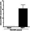
Figure 2. Real-time PCR data showing quantification of delivered BMP7 gene copies in rabbit corneas after a single 5 min topical application of PEI2-GNPs-BMP7 transfection solution.
Figure 3
Figure 3. Representative images showing immunofluorescence staining for αSMA (green), a myofibroblast marker, in the stroma of laser-ablated corneal tissue sections obtained from rabbit eyes that received a single 5 min topical application of transfection solution; PEI2-GNPs naked plasmid (A) or BMP7 expressing plasmid (B).
Scale bar denotes 100 µm.
Figure 4
Figure 4. Representative images showing immunofluorescence staining for fibronectin (red) in the stroma of laser ablated corneal tissue sections obtained from rabbit eyes that received a single 5 min topical application of transfection solution made of PEI2-GNPs naked plasmid (A) or BMP7 expressing plasmid (B).
Scale bar denotes 100 µm.
Figure 5
Figure 5. Quantification of corneal haze in live animals (A), and SMA+ (B) and fibronectin+ (C) cells in corneal tissues 4-week after laser ablation and +/− BMP7 gene transfer.
PEI2-GNPs mediated BMP7 gene transfer significantly reduced (p<0.05) in corneal haze, and decreased SMA+ cells (p<0.001) and fibronectin-immunostained area (p<0.01).
Figure 6
Figure 6. Representative images showing TUNEL assay data of rabbit corneas collected 4 weeks after laser ablation and one 5 min topical application of PEI2-GNPs naked plasmid (A) or BMP7 plasmid (B) transfection solution.
Tissue sections of rabbit corneas transfected with naked plasmid (A) or BMP7 plasmid (B) depicted no significant difference in TUNEL+ cells between the two groups. As expected many TUNEL+ cells in corneal epithelium were observed due to normal epithelium turnover. Scale bar denotes 100 µm.
Figure 7
Figure 7. Representative images showing CD11b immunostaining in rabbit corneas collected 4 weeks after laser ablation without (A) and with BMP7 (B) gene transfer.
No significant difference in the CD11b+ cells in tissue sections of rabbit corneas transfected with PEI2-GNPs naked (A) or BMP7 (B) plasmid was detected. Scale bar denotes 100 µm.
Figure 8
Figure 8. Representative osteocalcin immunofluorescence of rabbit corneas collected 4 weeks after laser ablation and PEI2-GNPs naked (A) or BMP7-expressing plasmid (B) transfection solution treatment, and tissue sections of horse hoof with laminitis (C).
Neither un-transfected nor BMP7-transfected rabbit corneas showed osteocalcin+ cells. This suggests that localized BMP7 overexpression in the cornea does not cause osteoblast recruitment. The horse hoof of laminitis, positive control, showed many osteocalcin+ cells (C). Scale bar denotes 100 µm.
Figure 9
Figure 9. Representative alizarin red (A-C) and vonKossa (D-F) staining in rabbit corneas and horse hoof laminitis tissue sections.
Rabbit corneas collected 4 weeks after laser ablation and PEI2-GNPs naked (A, D) or BMP7-expressing plasmid (B, E) treatment showed no alizarin red (A, B) or vonKossa (D, E) staining. This suggests that localized BMP7 gene transfer in rabbit cornea does not cause calcium deposits. Positive controls of horse hoof laminitis tissues (C, F) showed strong alizarin red (C) and vonKossa (F) staining. Scale bar denotes 100 µm.
Figure 10
Figure 10. Representative immunofluorescence image showing pSmad-1/5/8 (Red) staining in HCFs transfected with PEI2-GNPs naked plasmid (A–C) or BMP7-expressing plasmid (D–F).
BMP7 overexpressing HCFs showed significantly higher pSmad-1/5/8 nuclear localization (C; 88±5%; p<0.0001). Nuclei are stained blue. Scale bar denotes 100 µm.
Figure 11
Figure 11. Representative western blotting demonstrating pSmad1/5/8, Smad6 and αSMA protein levels in HCFs transfected with PEI2-GNPs naked plasmid or BMP7-expressing plasmid.
BMP7-transfected HCFs grown in the presence of TGFβ demonstrated a statistically significant increased pSmad-1/5/8 (95%; p<0.001) and Smad6 (53%, p<0.001), and decreased αSMA (78%; p<0.001) protein levels.

Similar articles

Cited by

References

    1. Mohan RR, Tovey JC, Sharma A, Tandon A (2012) Gene therapy in the cornea: 2005–present. Prog Retin Eye Res 31: 43–64. - PMC - PubMed
    1. Nien CJ, Flynn KJ, Chang M, Brown D, Jester JV (2011) Reducing peak corneal haze after photorefractive keratectomy in rabbits: prednisolone acetate 1.00% versus cyclosporine A 0.05%. J Cataract Refract Surg 37: 937–944. - PMC - PubMed
    1. Srinivasan M, Mascarenhas J, Rajaraman R, Ravindran M, Lalitha P, et al. (2012) Steroids for Corneal Ulcers Trial Group. Corticosteroids for bacterial keratitis: the Steroids for Corneal Ulcers Trial (SCUT). Arch Ophthalmol 130: 143–150. - PMC - PubMed
    1. Bedei A, Marabotti A, Giannecchini I, Ferretti C, Montagnani M, et al. (2006) Photorefractive keratectomy in high myopic defects with or without intraoperative mitomycin C: 1-year results. Eur J Ophthalmol 16: 229–234. - PubMed
    1. Camellin M (2004) Laser epithelial keratomileusis with mitomycin C: indications and limits. J Refract Surg 20: S693–S698. - PubMed

Publication types

MeSH terms