

Evaluation of small intestine grafts decellularization methods for corneal tissue engineering
- PMID: 23799114
- PMCID: PMC3682956
- DOI: 10.1371/journal.pone.0066538
Evaluation of small intestine grafts decellularization methods for corneal tissue engineering
Abstract
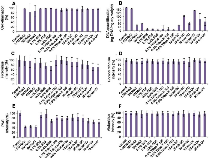
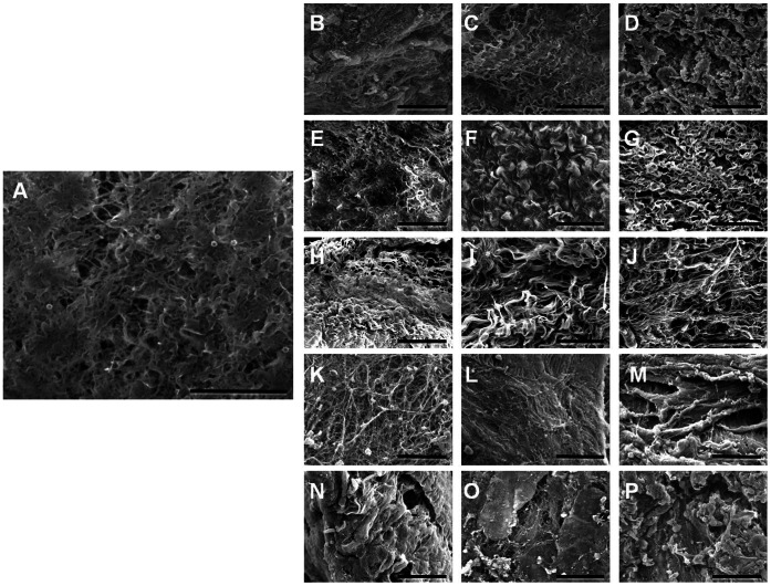
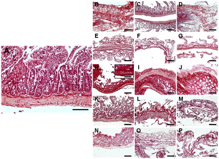
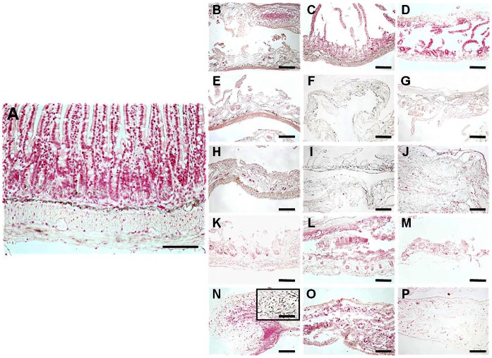
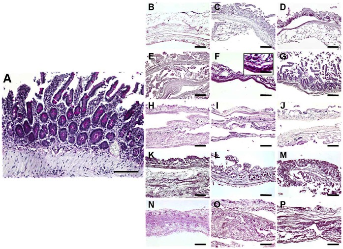
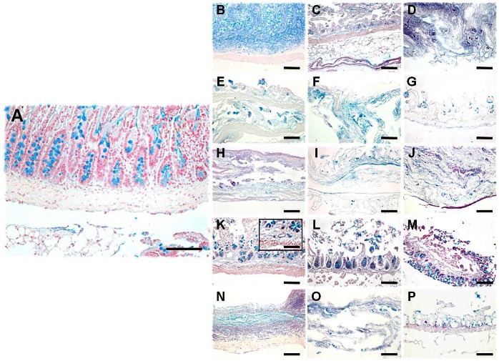
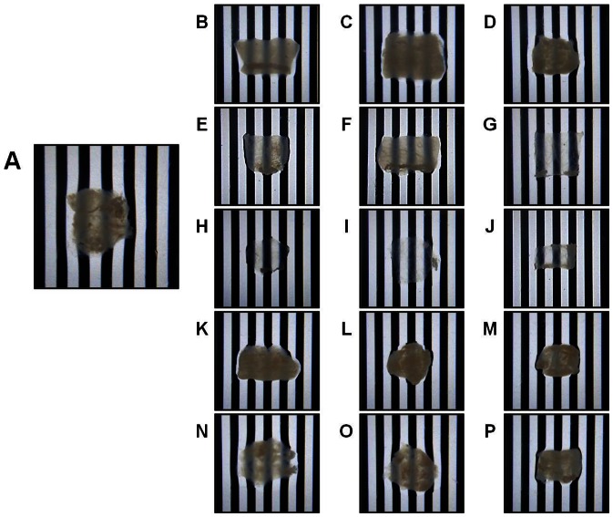
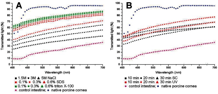
Advances in the development of cornea substitutes by tissue engineering techniques have focused on the use of decellularized tissue scaffolds. In this work, we evaluated different chemical and physical decellularization methods on small intestine tissues to determine the most appropriate decellularization protocols for corneal applications. Our results revealed that the most efficient decellularization agents were the SDS and triton X-100 detergents, which were able to efficiently remove most cell nuclei and residual DNA. Histological and histochemical analyses revealed that collagen fibers were preserved upon decellularization with triton X-100, NaCl and sonication, whereas reticular fibers were properly preserved by decellularization with UV exposure. Extracellular matrix glycoproteins were preserved after decellularization with SDS, triton X-100 and sonication, whereas proteoglycans were not affected by any of the decellularization protocols. Tissue transparency was significantly higher than control non-decellularized tissues for all protocols, although the best light transmittance results were found in tissues decellularized with SDS and triton X-100. In conclusion, our results suggest that decellularized intestinal grafts could be used as biological scaffolds for cornea tissue engineering. Decellularization with triton X-100 was able to efficiently remove all cells from the tissues while preserving tissue structure and most fibrillar and non-fibrillar extracellular matrix components, suggesting that this specific decellularization agent could be safely used for efficient decellularization of SI tissues for cornea TE applications.
Conflict of interest statement
Figures
Similar articles
-
Fast, robust and effective decellularization of whole human livers using mild detergents and pressure controlled perfusion.Mater Sci Eng C Mater Biol Appl. 2020 Mar;108:110200. doi: 10.1016/j.msec.2019.110200. Epub 2019 Sep 12. Mater Sci Eng C Mater Biol Appl. 2020. PMID: 31923991
-
Combination of freeze-thaw with detergents: A promising approach to the decellularization of porcine carotid arteries.Biomed Mater Eng. 2019;30(2):191-205. doi: 10.3233/BME-191044. Biomed Mater Eng. 2019. PMID: 30741667
-
Triton X-100 combines with chymotrypsin: A more promising protocol to prepare decellularized porcine carotid arteries.Biomed Mater Eng. 2017;28(5):531-543. doi: 10.3233/BME-171694. Biomed Mater Eng. 2017. PMID: 28854493
-
Decellularization and recellularization of cornea: Progress towards a donor alternative.Methods. 2020 Jan 15;171:86-96. doi: 10.1016/j.ymeth.2019.05.009. Epub 2019 May 22. Methods. 2020. PMID: 31128238 Review.
-
Decellularization methods for developing porcine corneal xenografts and future perspectives.Xenotransplantation. 2019 Nov;26(6):e12564. doi: 10.1111/xen.12564. Epub 2019 Oct 28. Xenotransplantation. 2019. PMID: 31659811 Free PMC article. Review.
Cited by
-
Do cells contribute to tendon and ligament biomechanics?PLoS One. 2014 Aug 15;9(8):e105037. doi: 10.1371/journal.pone.0105037. eCollection 2014. PLoS One. 2014. PMID: 25126746 Free PMC article.
-
Decellularization of Articular Cartilage: A Hydrochloric Acid-Based Strategy.Methods Mol Biol. 2023;2598:301-311. doi: 10.1007/978-1-0716-2839-3_21. Methods Mol Biol. 2023. PMID: 36355300
-
Preconditioning process for dermal tissue decellularization using electroporation with sonication.Regen Biomater. 2021 Dec 2;9(1):rbab071. doi: 10.1093/rb/rbab071. eCollection 2022. Regen Biomater. 2021. PMID: 35449827 Free PMC article.
-
Matrix bound nanovesicle-associated IL-33 activates a pro-remodeling macrophage phenotype via a non-canonical, ST2-independent pathway.J Immunol Regen Med. 2019 Mar;3:26-35. doi: 10.1016/j.regen.2019.01.001. Epub 2019 Feb 1. J Immunol Regen Med. 2019. PMID: 31656879 Free PMC article.
-
Expression of Basement Membrane Molecules by Wharton Jelly Stem Cells (WJSC) in Full-Term Human Umbilical Cords, Cell Cultures and Microtissues.Cells. 2023 Feb 15;12(4):629. doi: 10.3390/cells12040629. Cells. 2023. PMID: 36831296 Free PMC article.
References
-
- Langer R, Vacanti JP (1993) Tissue engineering. Science 260: 920–926. - PubMed
-
- Alaminos M, Del Carmen Sanchez-Quevedo M, Munoz-Avila JI, Serrano D, Medialdea S, et al. (2006) Construction of a complete rabbit cornea substitute using a fibrin-agarose scaffold. Invest Ophthalmol Vis Sci 47: 3311–3317. - PubMed
-
- Ionescu AM, de la Cruz Cardona J, Gonzalez-Andrades M, Alaminos M, Campos A, et al. (2010) UV absorbance of a bioengineered corneal stroma substitute in the 240–400 nm range. Cornea 29: 895–898 doi: 10.1097/ICO.0b013e3181ca3650 - DOI - PubMed
-
- Ionescu AM, Alaminos M, de la Cruz Cardona J, de Dios Garcia-Lopez Duran J, Gonzalez-Andrades M, et al. (2011) Investigating a novel nanostructured fibrin-agarose biomaterial for human cornea tissue engineering: rheological properties. J Mech Behav Biomed Mater 4: 1963–1973 doi: 10.1016/j.jmbbm.2011.06.013 - DOI - PubMed
-
- Cardona Jde L, Ionescu AM, Gomez-Sotomayor R, Gonzalez-Andrades M, Campos A, et al. (2011) Transparency in a fibrin and fibrin-agarose corneal stroma substitute generated by tissue engineering. Cornea 30: 1428–1435 doi: 10.1097/ICO.0b013e31821bdfd4 - DOI - PubMed
Publication types
MeSH terms
Substances
LinkOut - more resources
Full Text Sources
Other Literature Sources
