. 2013 Jul 31;32(15):2172-85.
doi: 10.1038/emboj.2013.148. Epub 2013 Jun 25.

DNA polymerase κ-dependent DNA synthesis at stalled replication forks is important for CHK1 activation

Affiliations

DNA polymerase κ-dependent DNA synthesis at stalled replication forks is important for CHK1 activation

Rémy Bétous et al. EMBO J. .

Abstract

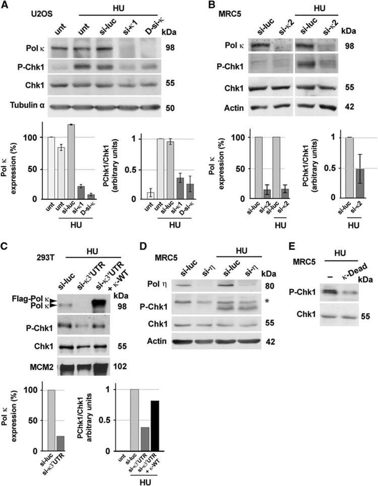
Formation of primed single-stranded DNA at stalled replication forks triggers activation of the replication checkpoint signalling cascade resulting in the ATR-mediated phosphorylation of the Chk1 protein kinase, thus preventing genomic instability. By using siRNA-mediated depletion in human cells and immunodepletion and reconstitution experiments in Xenopus egg extracts, we report that the Y-family translesion (TLS) DNA polymerase kappa (Pol κ) contributes to the replication checkpoint response and is required for recovery after replication stress. We found that Pol κ is implicated in the synthesis of short DNA intermediates at stalled forks, facilitating the recruitment of the 9-1-1 checkpoint clamp. Furthermore, we show that Pol κ interacts with the Rad9 subunit of the 9-1-1 complex. Finally, we show that this novel checkpoint function of Pol κ is required for the maintenance of genomic stability and cell proliferation in unstressed human cells.


Conflict of interest statement

The authors declare that they have no conflict of interest.

Figures

Figure 1
Figure 1
Pol κ is required for Chk1 phosphorylation after replication stress in human cells. (A, B) Western blot analysis of cell extracts from Human U2OS and MRC5 untransfected (unt) or transfected with control luciferase siRNA (si-luc), Pol κ siRNA (si-κ1, si-κ2, D-si-κ), untreated or treated with HU (2 mM, 3 h). Cell extracts were fractionated and soluble fractions were analysed with the indicated antibodies. Quantification was performed by ImageJ software; means±s.d. were presented. (C) Western blot analysis of whole cell extracts prepared from 293T cells treated with HU (2 mM, 3 h) transfected by si-RNA alone (si-luc or si-κ3’UTR) or co-transfected by si-κ3′UTR and a vector expressing FLAG-tagged wild-type Pol κ (κ-WT). (D, E) Pol η depletion with Pol η siRNA (si-η) or transfection with vector expressing the FLAG-tagged catalytic inactive mutant of Pol κ (Pol κ-Dead) was performed in MRC5 cells untreated or treated with HU (2 mM, 3 h). Source data for this figure is available on the online supplementary information page.
Figure 2
Figure 2
Pol κ depletion from Xenopus egg extracts affects Chk1 phosphorylation. (A) Xenopus egg extracts were either mock depleted (ΔMock) or depleted with an anti-XPol κ-specific antibody (ΔXpol κ) as described in Materials and methods. Egg supernatants after depletion were analysed by western blotting with the indicated antibodies. (B) Analysis of Chk1P-S344 in mock-depleted or XPol κ-depleted extracts treated with (+) or without (−) aphidicolin (15 μM) and reconstituted with 25 ng of recombinant Pol κ (R-Pol κ) performed as in B. Chk1 was used as the loading control. (C) Kinetics of DNA synthesis of mock-depleted (ΔMock; left panel) or XPol κ-depleted (ΔXPol κ; right panel) egg extracts in both the presence (+) or absence (−) of UV irradiation (800 J/m2) and absence (−) or presence of 5 mM caffeine. The extent of DNA synthesis of both extracts is expressed as percent (%) of the input DNA added to egg extracts at time=0. Source data for this figure is available on the online supplementary information page.
Figure 3
Figure 3
Pol κ is required for the accumulation of small nascent DNA in response to replication stress in Xenopus egg extracts. (A) The abundance of checkpoint factors in extracts Mock-depleted (ΔMock) or XPol κ-depleted (ΔXPol κ) was analysed by immunoblotting. (B) Chromatin binding of RPA in egg extracts mock depleted or depleted with XPol κ antibodies in the absence (−) or presence (+) of aphidicolin. (C) Sperm chromatin was replicated in egg extracts mock depleted (ΔMock) or depleted with XPol κ antibodies (ΔXPol κ) containing 15 μM aphidicolin (APH) and α-[P32]dCTP. At 40, 60 and 90 min, total DNA was purified as described (Van et al, 2010), replication intermediates were fractionated by denaturing polyacrylamide gel electrophoresis and detected by autoradiography after exposure to a PhosphoImager screen (Molecular Dynamics). Abundance of 25–150 nt long DNA intermediates was quantified by densitometric scanning and analysed with ImageJ software. (D) Sperm chromatin was incubated in control (ΔMock) or XPol κ (ΔXPol κ)-depleted egg extracts in the presence of aphidicolin (15 μM) for 60 min. Chromatin fractions were analysed by immunoblotting with the indicated antibodies. Western blot signals were quantified with ImageJ software, normalized to histone H3 signals. (E) Analysis of XPol κ chromatin binding. Demembranated sperm nuclei (2000, nuclei/μl of egg extract) were incubated in egg extracts for 90 min in the presence (+) or absence (−) of 15 or 750 μM aphidicolin. Chromatin fractions were obtained as described in Materials and methods and analysed by immunoblotting with the indicated antibodies. Source data for this figure is available on the online supplementary information page.
Figure 4
Figure 4
Pol κ suppresses formation of single-stranded DNA induced by replicative stress upon recovery from an HU block. (A, B) Persistence of BrdU-positive cells in Pol κ-deficient cells 3 h after release from HU. Exponentially growing MRC5 cells were transfected with the indicated si-RNA; 48 h later cells were treated with 2 mM HU for 3 h and collected (R0) or released for 3 h in fresh medium (R3). Detection of BrdU (green) and PCNA (red) was performed in non-denaturating conditions. DNA was counterstained with DAPI. IgG1/488 and -/555 were controls of immunodetections. Examples of negative and positive cells for BrdU labelling at R0 and R3 are shown in A; scale bar 10 μm. (B) At least 150 MRC5 were randomly acquired with wide field microscopy and BrdU-positive nuclei in PCNA-positive nuclei were quantified in three independent experiments. Standard deviations were indicated by error bars and a t-test was applied (*P<0.05). (C) Increased level of Ub-PCNA onto chromatin after HU in Pol κ-deficient cells. Extracts of MRC5 cells transfected with si-luc or si-κ1 were fractionated. Soluble and chromatin fractions were then subjected to immunoblotting with the indicated antibodies. Actinin and MCM7 served as loading controls for soluble and chromatin fractions, respectively. Source data for this figure is available on the online supplementary information page.
Figure 5
Figure 5
Pol κ depletion triggers perturbation of DNA replication program. (A) Increased RPA foci formation in nuclei from Pol κ-depleted cells. Left panel: MRC5 cells transfected with control siRNA (si-luc) or siRNAs targeting Pol κ (si-κ1) were randomly acquired (n>100 nuclei) by confocal microscopy and quantification of RPA-positive nuclei was performed 48 h after transfection; DNA content was visualized by DAPI staining. Right panel: cells transfected by the indicated siRNA were randomly acquired (n>80 cells) with wide field microscopy and the number of RPA foci per nucleus was automatically counted with ImageJ software; the distribution of the RPA foci per cell was presented and the P-value determined with the non-parametric Mann–Whitney test was 0.011 (*). (B) RPA accumulation onto chromatin in Pol κ-deficient cells. Extracts of MRC5 cells transfected with control siRNA (si-luc) or siRNAs targeting Pol κ (si-κ1) were fractionated. Chromatin and soluble fractions were then subjected to immunoblotting with the indicated antibodies. Extracts from the untransfected cells irradiated with UV (50 J/m2) serves as positive control for RPA hyperloading and Chk1 phosphorylation; actin serves as loading control. (C) Catalytic activity of Pol κ is required for preventing RPA accumulation onto chromatin in unstressed cells: western blot with the specified antibodies of chromatin cell extracts prepared from HeLa cells co-transfected by the indicated siRNA and empty vector (−) or vectors expressing FLAG-tagged wild-type Pol κ (Pol κ-WT) or the FLAG-tagged catalytic inactive mutant of Pol κ (Pol κ-Dead). (D) Depletion of Pol κ affects the spatial organization of active replication foci. Left panel: images of early, middle and late replication PCNA foci in U2OS cells analysed by immunofluorescence; scale bar: 10 μm. Right panel: quantification of MRC5 and HeLa cells with middle/late replication foci was performed (n>100 cells) for control (si-luc) and Pol κ-deficient cells (si-κ1, si-κ3′UTR) grown under 20 and 5% of oxygen. Source data for this figure is available on the online supplementary information page.
Figure 6
Figure 6
Increased 53BP1 nuclear bodies in G1 following Pol κ depletion. Untransfected (unt) or transfected with control siRNA (si-luc) or siRNAs targeting Pol κ (si-κ1, si-κ2) were untreated (AC) or treated with 0.2 μM of aphidicolin for 24 h (DF). 53PB1 was detected by immunofluorescence in G1 nuclei (cyclin A negative). Examples of positive nuclei for 53BP1 (red), Cyclin A (green) and DAPI (blue) staining are shown in A and D; B and E are representative experiments showing the distribution of the number of 53BP1 foci per cell in control and Pol κ-deficient cells (n=100); Box, 25–75 percentile range; whiskers, minimum and maximum values. Mann–Whitney test was applied to compare Pol κ-deficient cells data set with the control cells (***P<0.001). In (C) and (F), 53BP1-positive nuclei were scored and classified in the indicated categories based on the number of 53BP1 nuclear bodies. Experiments were done three times (n=100) and standard deviations are indicated by error bars.
Figure 7
Figure 7
Analysis of cell-cycle progression and cell proliferation in Pol κ-deficient cells. (A) The percentage (%) of G2/M cells was determined in HeLa or in HeLa XPAKD cells by flow cytometry analysis on control (si-luc) and Pol κ-deficient cells (si-κ1 and si-κ2). Experiments were done three times and standard deviations are indicated by error bars. P-values were calculated using Student’s t-test (*P<0.05; **P<0.01). (B) Evaluation of the function of the G2/M checkpoint: the relative mitotic index in Pol κ-deficient cells compared with control cells was measured by monitoring the phosphorylated form of Histone H3 by flow cytometry analysis; DNA content was determined after DNA labelling with propidium iodide (PI). The percentage of H3P-positive cells was determined in three independent experiments and standard deviations were indicated by error bars. P-values were calculated using Student’s t-test (*P<0.05; ***P<0.001). (C) Doubling time calculated from growth curve done during 96 h on control and Pol κ-deficient HeLa and MRC5 cells. Experiments were done three times in triplicate and standard deviations are indicated by error bars. P-values were calculated using Student’s t-test (*P<0.05; **P<0.01; ***P<0.001).
Figure 8
Figure 8
A model for the checkpoint function of Pol κ. Upon replication stress, new short RNA-DNA primers continue to be synthesized and elongated on the unwound ssDNA downstream of a stalled replisome. On the lagging strand, primers accumulate through recycling of Pol α and are elongated by Pol δ with PCNA. On the leading strand, Pol α could be also recruited and elongation of the primers could be made by the distributive Pol κ in order to allow high levels of 5’ ends primer-template junctions. Accumulation of short nascent DNAs on both strands results in multiple 5′ ends of primer-template junctions facilitating the binding of the 9-1-1 complex and subsequently checkpoint activation.

Comment in

  • Pol κ in replication checkpoint.
    Maiorano D, Hoffmann JS. Maiorano D, et al. Cell Cycle. 2013 Dec 15;12(24):3713-4. doi: 10.4161/cc.26976. Epub 2013 Oct 28. Cell Cycle. 2013. PMID: 24189533 Free PMC article. No abstract available.

References

    1. Avkin S, Goldsmith M, Velasco-Miguel S, Geacintov N, Friedberg EC, Livneh Z (2004) Quantitative analysis of translesion DNA synthesis across a benzo[a]pyrene-guanine adduct in mammalian cells: the role of DNA polymerase kappa. J Biol Chem 279: 53298–53305 - PubMed
    1. Bergoglio V, Boyer AS, Walsh E, Naim V, Legube G, Lee MY, Rey L, Rosselli F, Cazaux C, Eckert KA, Hoffmann JS (2013) DNA synthesis by Pol eta promotes fragile site stability by preventing under-replicated DNA in mitosis. J Cell Biol 201: 395–408 - PMC - PubMed
    1. Betous R, Rey L, Wang G, Pillaire MJ, Puget N, Selves J, Biard DS, Shin-ya K, Vasquez KM, Cazaux C, Hoffmann JS (2009) Role of TLS DNA polymerases eta and kappa in processing naturally occurring structured DNA in human cells. Mol Carcinog 48: 369–378 - PMC - PubMed
    1. Bi X, Slater DM, Ohmori H, Vaziri C (2005) DNA polymerase kappa is specifically required for recovery from the benzo[a]pyrene-dihydrodiol epoxide (BPDE)-induced S-phase checkpoint. J Biol Chem 280: 22343–22355 - PubMed
    1. Biard DS, Despras E, Sarasin A, Angulo JF (2005) Development of new EBV-based vectors for stable expression of small interfering RNA to mimick human syndromes: application to NER gene silencing. Mol Cancer Res 3: 519–529 - PubMed

Publication types