Review
. 2013 Jul;16(7):805-15.
doi: 10.1038/nn.3427. Epub 2013 Jun 25.

Targeting neurons and photons for optogenetics

Affiliations
Review

Targeting neurons and photons for optogenetics

Adam M Packer et al. Nat Neurosci. 2013 Jul.

Abstract

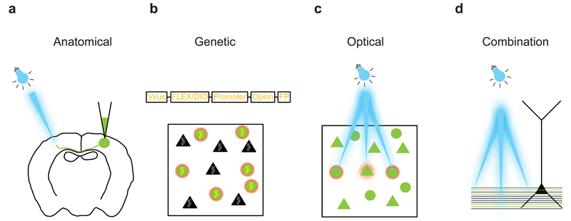
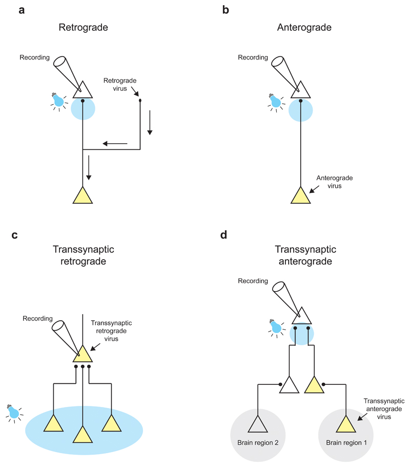
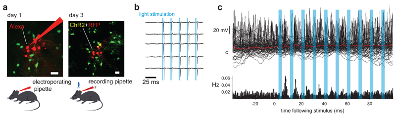
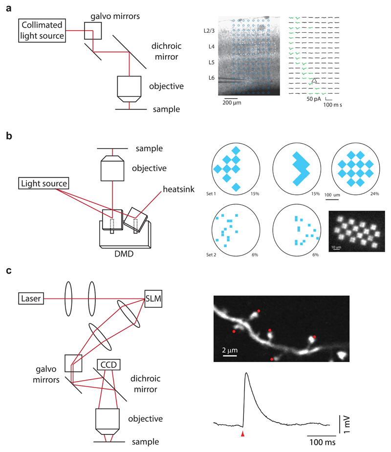
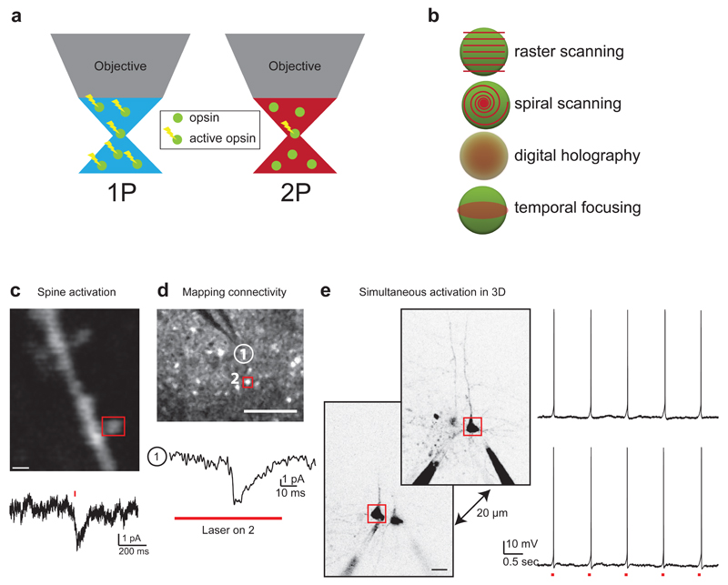
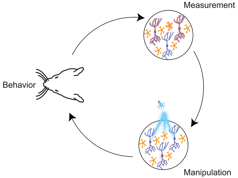
Optogenetic approaches promise to revolutionize neuroscience by using light to manipulate neural activity in genetically or functionally defined neurons with millisecond precision. Harnessing the full potential of optogenetic tools, however, requires light to be targeted to the right neurons at the right time. Here we discuss some barriers and potential solutions to this problem. We review methods for targeting the expression of light-activatable molecules to specific cell types, under genetic, viral or activity-dependent control. Next we explore new ways to target light to individual neurons to allow their precise activation and inactivation. These techniques provide a precision in the temporal and spatial activation of neurons that was not achievable in previous experiments. In combination with simultaneous recording and imaging techniques, these strategies will allow us to mimic the natural activity patterns of neurons in vivo, enabling previously impossible 'dream experiments'.


Figures

Figure 1
Figure 1. Intersectional strategies for targeting optogenetic manipulation
(a) Physical delivery of virus to a given anatomical location can exploit or uncover circuit connectivity patterns either by making use of axonal projections or by using viruses that are able to cross one or more synapses. (b) Addressing cell types can be performed if the cell type of interest has a known genetic identity. (c) Directing the illumination source to a given set of cells or even individual neurons and processes is useful when the targets of interest are separated in space relative to the spatial resolution of the technique employed. (d) These three strategies can be combined as shown in this example in which axons of a particular cell class projecting to a subcellular domain of a neuron are photostimulated at different distances from the neuron.
Figure 2
Figure 2. Viral targeting of optogenetic tools using knowledge of circuit connectivity
Schematic illustration of different strategies for targeting optogenetic tools to specific cell types based on their connectivity pattern. Neurons expressing an optogenetic tool are indicated in yellow; arrows next to cellular processes indicate the direction of viral spread; and the location of light stimulation is shown in blue. (a) Use of a retrograde virus with targeted virus injection to an axon projection region. (b) Use of an anterograde virus with targeted virus injection to the somatic region. (c) Use of a transsynaptic retrograde virus starting from virus introduction (or infection) of a single postsynaptic cell which leads to optogene expression in monosynaptically connected presynaptic partners. (d) Use of a transsynaptic anterograde virus starting from virus injection in a given brain region to cause optogene expression in synaptically connected downstream neurons.
Figure 3
Figure 3. Targeting optogene expression using single-cell electroporation
a, Left panel: Two-photon Z-stack projection of mouse cortex during successive single-cell electroporation; the transgenic mouse expresses GFP in GAD67-positive cortical inhibitory neurons (green cells). The red neurons have been electroporated with Alexa Fluor 594 (red cells) and plasmid DNA encoding RFP and ChR2. Right panel: 48 hours later the electroporated cells express RFP (red) suggesting that ChR2 is also expressed. b Cell-attached recording from a red cell confirms that blue light stimulation using an LED drives the neuron to fire an action potential precisely and reliably. c Whole-cell recording from a green cell (inhibitory, not electroporated) shows an increased firing probability following a light stimulus. The average membrane potential shows a depolarizing transient 5 ms after the onset of light stimulus, suggesting a direct synaptic connection from some of the electroporated neurons onto this cell. Unpublished data from M. London, L. Beeren and M. Hausser.
Figure 4
Figure 4. Patterned illumination strategies
(a) Left, pointing a single beam with galvanometer mirrors is the most straightforward implementation of directing a focused beam of light onto different locations within a sample. Right, this approach is particularly useful for mapping studies in which independent activation of small, localized subsets of labeled neurons or axons is desired for readout by downstream neurons. (b) Left, pointing multiple beams with a digital micromirror device. Right, this enables more complex patterns of activation across large areas of tissue, which has proven useful in studies of retinal circuitry and zebrafish behavior. (c) Left, creating holographic patterns with a spatial light modulator combines the power of generating multiple individual beamlets with high efficiency in directing power into those beamlets. Right, this enables multi-site activation, when combined with two-photon excitation (see Fig. 5).
Figure 5
Figure 5. One-photon versus two-photon activation strategies: from spines to circuits
(a) In one-photon excitation (left), all opsin molecules illuminated above and below the focal plane of interest are excited. Alternatively, in two-photon excitation (right), generally only opsin molecules in the focal plane are excited (but see Ref. 76), leading to optical sectioning that allows activation to be restricted to the particular neurons of interest. (b) Spatiotemporal patterns for illuminating neurons with two-photon beams require different power budgets and yield different spatial and temporal resolutions (see Table 1). (c) Two-photon point stimulation of a dendritic spine on a neuron expressing C1V1 (top panel, scale bar 1 um) generates current detectable at the soma (bottom trace). (d) Two-photon raster-scanning of neuron 2 (top panel, red box) during electrophysiological recording from neuron 1 (white circle, top panel and bottom trace) indicates that neuron 2 is monosynaptically connected to neuron 1 (scale bar 100 µm). (e) Simultaneous action potential generation in two neurons in 3D using a spatial light modulator to generate separate laser beamlets over each neuron (scale bar 20 µm). Data in panels c,d,e adapted from Ref. 84.
Figure 6
Figure 6. Using targeted optogenetics to enable ‘dream experiments’.
A schematic illustration of how ‘targeted optogenetics’ can be used to probe the neural code in a cortical circuit. The figure highlights the close interplay that is necessary between behavioural experiments, optical readout of patterns of activity, and replay of the same patterns in the ‘right’ neurons using optogenetics. Targeted optogenetics allows the precision of temporal patterns, and the precise membership of the neuronal ensemble, to be tested directly to investigate their importance for the neural code driving the behaviour.

References

    1. Miesenbock G. The optogenetic catechism. Science. 2009;326:395–399. doi: 10.1126/science.1174520. - DOI - PubMed
    1. Bamann C, Nagel G, Bamberg E. Microbial rhodopsins in the spotlight. Curr Opin Neurobiol. 2010;20:610–616. doi: 10.1016/j.conb.2010.07.003. - DOI - PubMed
    1. Yizhar O, Fenno LE, Davidson TJ, Mogri M, Deisseroth K. Optogenetics in neural systems. Neuron. 2011;71:9–34. doi: 10.1016/j.neuron.2011.06.004. - DOI - PubMed
    1. Madisen L, et al. A toolbox of Cre-dependent optogenetic transgenic mice for light-induced activation and silencing. Nat Neurosci. 2012;15:793–802. doi: 10.1038/nn.3078. - DOI - PMC - PubMed
    1. Kienle E, et al. Engineering and evolution of synthetic adeno-associated virus (AAV) gene therapy vectors via DNA family shuffling. Journal of visualized experiments : JoVE. 2012 doi: 10.3791/3819. - DOI - PMC - PubMed

Publication types