Review
. 2012 Sep-Oct;1(5):609-28.
doi: 10.1002/wdev.44. Epub 2012 Mar 14.

The endocrine pancreas: insights into development, differentiation, and diabetes

Affiliations
Review

The endocrine pancreas: insights into development, differentiation, and diabetes

Teresa L Mastracci et al. Wiley Interdiscip Rev Dev Biol. 2012 Sep-Oct.

Abstract

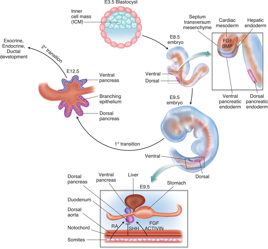
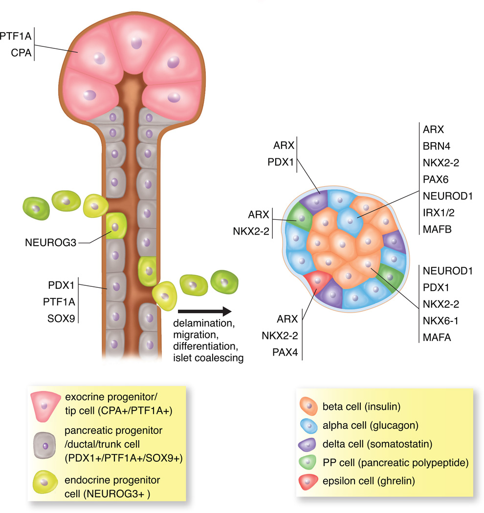
In the developing embryo, appropriate patterning of the endoderm fated to become pancreas requires the spatial and temporal coordination of soluble factors secreted by the surrounding tissues. Once pancreatic progenitor cells are specified in the developing gut tube epithelium, epithelial-mesenchymal interactions, as well as a cascade of transcription factors, subsequently delineate three distinct lineages, including endocrine, exocrine, and ductal cells. Simultaneous morphological changes, including branching, vascularization, and proximal organ development, also influence the process of specification and differentiation. Decades of research using mouse genetics have uncovered many of the key factors involved in pancreatic cell fate decisions. When pancreas development or islet cell functions go awry, due to mutations in genes important for proper organogenesis and development, the result can lead to a common pancreatic affliction, diabetes mellitus. Current treatments for diabetes are adequate but not curative. Therefore, researchers are utilizing the current understanding of normal embryonic pancreas development in vivo, to direct embryonic stem cells toward a pancreatic fate with the goal of transplanting these in vitro generated 'islets' into patients. Mimicking development in vitro has proven difficult; however, significant progress has been made and the current differentiation protocols are becoming more efficient. The continued partnership between developmental biologists and stem cell researchers will guarantee that the in vitro generation of insulin-producing β cells is a possible therapeutic option for the treatment of diabetes.


Figures

Figure 1
Figure 1. Pancreatic endoderm patterning and specification
The portion of the endoderm fated to become dorsal and ventral pancreas is patterned and specified due to the influence of secreted molecules and signalling pathways from adjacent tissues and cells. The influence of FGF and BMP signalling from the cardiac mesoderm and septum transversum mesenchyme, respectively, affect the patterning of ventral pancreatic endoderm. Retinoic acid (RA), SHH, FGF and ACTIVIN signalling from the paraxial mesoderm and notochord allow for patterning of the dorsal pancreatic endoderm. The stage from E9.5 through E12.5 marks the primary transition. During this period, the pancreatic progenitor cells are specified, first wave endocrine cells are present, and epithelial branching morphogenesis begins. The secondary transition occurs after E12.5 and is marked by a major wave of endocrine cell specification and development, as well as further ductal morphogenesis and exocrine lineage differentiation.
Figure 2
Figure 2. Pancreatic specification is unaltered in embryos lacking either Pdx1 or Ptf1a
Sagittal sections from E10.5 Pdx1 null (A) and wildtype littermate (B), as well as Ptf1a null (C) and wildtype littermate (D) embryos were stained by immunofluorescence with antibodies against the transcription factor FOXA (demarking endoderm) and the hormone glucagon (identifying the pancreatic domain). In both null embryos, the region fated to become the dorsal pancreas (dp) is present, contains glucagon-expressing cells, and appears similar in both the null and wildtype. stm, septum transversum mesenchyme. DAPI marks all nuclei. 20X
Figure 3
Figure 3. Pancreatic and biliary domains in the wildtype E10.5 embryo
Adjacent sagittal sections from E10.5 wildtype embryo, stained by immunofluorescence with antibodies against FOXA (A), and PDX1, SOX17 (B). The domains of the dorsal pancreas (dp), ventral pancreas (vp), biliary domain (bil) and septum transversum mesenchyme (stm) are noted. While all domains express FOXA (A), PDX1+ cells are found only in the pancreatic domains, and are distinct from the SOX17+ cells in the biliary domain (B). DAPI marks all nuclei. 20X
Figure 4
Figure 4. Early “first wave” glucagon-expressing alpha cells
Early “first wave” alpha cells are present in mouse models despite the deletion of factors necessary for pancreas development and endocrine specification. Sagittal sections of E10.5 embryos from a representative wildtype littermate (A), Pdx1 null (B), Ptf1a null (C), Neurog3 null (D), Nkx2-2 null (E), and pancreas-specific deletion of Arx (F), were stained by immunofluorescence for the hormone glucagon (A–F). In all images glucagon-expressing cells are present. All images are of the dorsal pancreas. DAPI marks all nuclei. 40X
Figure 5
Figure 5. Islet cell specification in the developing pancreas
Multipotent pancreatic progenitor cells (PDX1+/PTF1A+/SOX9+) are present in the ductal epithelium in the developing pancreas. In addition, multipotent CPA+/PTF1A+ cells at the tips in early development are multipotent progenitor cells but later in development are restricted to differentiating into only the exocrine lineage. All hormone-producing endocrine cell lineages are derived from the endocrine progenitor cells (NEUROG3+), which delaminate from the ductal epithelium. Endocrine progenitors can differentiate into all five hormone-expressing cell types including insulin-producing beta cells, glucagon-producing alpha cells, somatostatin-producing delta cells, pancreatic polypeptide-producing PP cells and ghrelin-producing epsilon cells. As development proceeds, these differentiated hormone+ cells will coalesce and form the islets of Langerhans. The influence of various transcription factors determines the specific endocrine cell produced.

References

    1. Murtaugh LC, Leach SD. A case of mistaken identity? Nonductal origins of pancreatic "ductal" cancers. Cancer Cell. 2007;11:211–213. - PubMed
    1. Pour PM, Pandey KK, Batra SK. What is the origin of pancreatic adenocarcinoma? Mol Cancer. 2003;2:13. - PMC - PubMed
    1. Oberg K. Pancreatic endocrine tumors. Semin Oncol. 2010;37:594–618. - PubMed
    1. Zorn AM, Wells JM. Vertebrate endoderm development and organ formation. Annu Rev Cell Dev Biol. 2009;25:221–251. - PMC - PubMed
    1. Chen Y, Pan FC, Brandes N, Afelik S, Solter M, Pieler T. Retinoic acid signaling is essential for pancreas development and promotes endocrine at the expense of exocrine cell differentiation in Xenopus. Dev Biol. 2004;271:144–160. - PubMed

Publication types