

The Drosophila midgut: a model for stem cell driven tissue regeneration
- PMID: 23799573
- PMCID: PMC5489342
- DOI: 10.1002/wdev.51
The Drosophila midgut: a model for stem cell driven tissue regeneration
Abstract
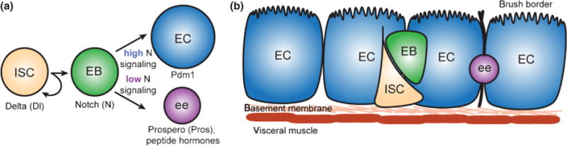
The Drosophila and mammalian digestive systems bear striking similarities in genetic control and cellular composition, and the Drosophila midgut has emerged as an amenable model for dissecting the mechanisms of tissue homeostasis. The Drosophila midgut is maintained by multipotent intestinal stem cells (ISCs) that give rise to all cell types in the intestinal epithelium and are required for long-term tissue homeostasis. ISC proliferation rate increases in response to a myriad of chemical and bacterial insults through the release of JAK-STAT and EGFR ligands from dying enterocytes that activate the JAK-STAT and EGFR pathways in ISCs. The Hippo and JNK pathways converge upon JAK-STAT and EGFR signaling, presumably in response to specific stresses, and JNK and insulin signaling have been shown to be critical in response to age-related stresses. This review details these emerging mechanisms of tissue homeostasis and the proliferative response of ISCs to epithelial damage, environmental stresses, and aging.
Copyright © 2012 Wiley Periodicals, Inc.
Figures

References
-
- Ohlstein B, Spradling A. The adult Drosophila posterior midgut is maintained by pluripotent stem cells. Nature. 2006;439:470–474. - PubMed
-
- Micchelli CA, Perrimon N. Evidence that stem cells reside in the adult Drosophila midgut epithelium. Nature. 2006;439:475–479. - PubMed
-
- Ohlstein B, Spradling A. Multipotent Drosophila intestinal stem cells specify daughter cell fates by differential Notch signaling. Science. 2007;315:988–992. - PubMed
MeSH terms
Substances
Grants and funding
LinkOut - more resources
Full Text Sources
Medical
Molecular Biology Databases
Research Materials
Miscellaneous
