

The etiology and molecular genetics of human pigmentation disorders
- PMID: 23799582
- PMCID: PMC3694277
- DOI: 10.1002/wdev.72
The etiology and molecular genetics of human pigmentation disorders
Abstract
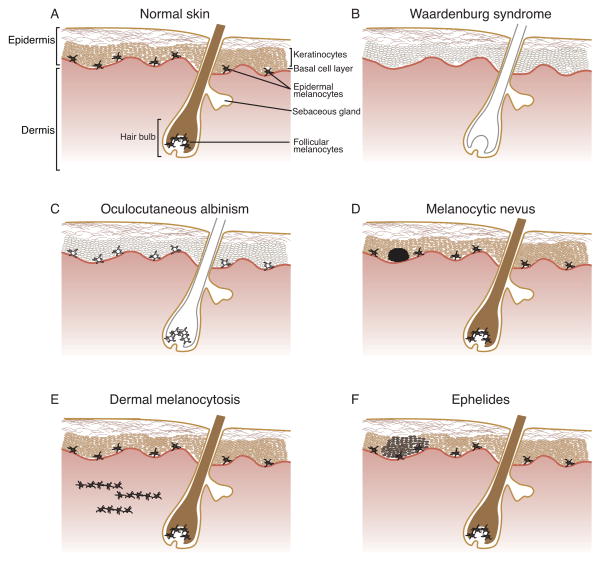
Pigmentation, defined as the placement of pigment in skin, hair, and eyes for coloration, is distinctive because the location, amount, and type of pigmentation provides a visual manifestation of genetic heterogeneity in pathways regulating the pigment-producing cells, melanocytes. The scope of this genetic heterogeneity in humans ranges from normal to pathological pigmentation phenotypes. Clinically, normal human pigmentation encompasses a variety of skin and hair color as well as punctate pigmentation such as melanocytic nevi (moles) or ephelides (freckles), while abnormal human pigmentation exhibits markedly reduced or increased pigment levels, known as hypopigmentation and hyperpigmentation, respectively. Elucidation of the molecular genetics underlying pigmentation has revealed genes important for melanocyte development and function. Furthermore, many pigmentation disorders show additional defects in cells other than melanocytes, and identification of the genetic insults in these disorders has revealed pleiotropic genes, where a single gene is required for various functions in different cell types. Thus, unravelling the genetics of easily visualized pigmentation disorders has identified molecular similarities between melanocytes and less visible cell types/tissues, arising from a common developmental origin and/or shared genetic regulatory pathways. Herein we discuss notable human pigmentation disorders and their associated genetic alterations, focusing on the fact that the developmental genetics of pigmentation abnormalities are instructive for understanding normal pathways governing development and function of melanocytes.
Copyright © 2012 Wiley Periodicals, Inc.
Conflict of interest statement
The authors declare no conflicts of interest
Figures
References
-
- Thomas I, Kihiczak GG, Fox MD, Janniger CK, Schwartz RA. Piebaldism: an update. Int J Dermatol. 2004;43:716–719. - PubMed
-
- Thomas AJ, Erickson CA. The making of a melanocyte: the specification of melanoblasts from the neural crest. Pigment cell & melanoma research. 2008;21:598–610. - PubMed
-
- Adameyko I, Lallemend F, Aquino JB, Pereira JA, Topilko P, Müller T, Fritz N, Beljajeva A, Mochii M, Liste I, Usoskin D, Suter U, Birchmeier C, Ernfors P. Schwann cell precursors from nerve innervation are a cellular origin of melanocytes in skin. Cell. 2009;139:366–379. - PubMed
-
- Luciani F, Champeval D, Herbette A, Denat L, Aylaj B, Martinozzi S, Ballotti R, Kemler R, Goding CR, De Vuyst F, Larue L, Delmas V. Biological and mathematical modeling of melanocyte development. Development. 2011;138:3943–3954. - PubMed
Further Reading/Resources
-
- Nordlund JJ, Boissy RE, Hearing VJ, King RA, Oetting WS, Ortonne JP. The Pigmentary System: Physiology and Pathophysiology. Blackwell Publishing; Malden, Massachusetts: 2006.
-
-
The Online Metabolic and Molecular Bases of Inherited Disease
-
-
-
Online Mendelian Inheritance in Man
-
Publication types
MeSH terms
Substances
Grants and funding
LinkOut - more resources
Full Text Sources
Medical
