Synthesis of CdTe quantum dot-conjugated CC49 and their application for in vitro imaging of gastric adenocarcinoma cells
- PMID: 23800369
- PMCID: PMC3695781
- DOI: 10.1186/1556-276X-8-294
Synthesis of CdTe quantum dot-conjugated CC49 and their application for in vitro imaging of gastric adenocarcinoma cells
Abstract
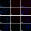
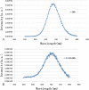
The purpose of this experiment was to investigate the visible imaging of gastric adenocarcinoma cells in vitro by targeting tumor-associated glycoprotein 72 (TAG-72) with near-infrared quantum dots (QDs). QDs with an emission wavelength of about 550 to 780 nm were conjugated to CC49 monoclonal antibodies against TAG-72, resulting in a probe named as CC49-QDs. A gastric adenocarcinoma cell line (MGC80-3) expressing high levels of TAG-72 was cultured for fluorescence imaging, and a gastric epithelial cell line (GES-1) was used for the negative control group. Transmission electron microscopy indicated that the average diameter of CC49-QDs was 0.2 nm higher compared with that of the primary QDs. Also, fluorescence spectrum analysis indicated that the CC49-QDs did not have different optical properties compared to the primary QDs. Immunohistochemical examination and in vitro fluorescence imaging of the tumors showed that the CC49-QDs probe could bind TAG-72 expressed on MGC80-3 cells.
Figures


Similar articles
-
In vitro gastric cancer cell imaging using near-infrared quantum dot-conjugated CC49.Oncol Lett. 2012 Nov;4(5):996-1002. doi: 10.3892/ol.2012.870. Epub 2012 Aug 20. Oncol Lett. 2012. PMID: 23162639 Free PMC article.
-
A fast synthesis of near-infrared emitting CdTe/CdSe quantum dots with small hydrodynamic diameter for in vivo imaging probes.Nanoscale. 2011 Nov;3(11):4724-32. doi: 10.1039/c1nr10933b. Epub 2011 Oct 11. Nanoscale. 2011. PMID: 21989776
-
[Quantitative determination of pazufloxacin using water-soluble quantum dots as fluorescent probes].Guang Pu Xue Yu Guang Pu Fen Xi. 2008 Jun;28(6):1317-21. Guang Pu Xue Yu Guang Pu Fen Xi. 2008. PMID: 18800713 Chinese.
-
Synthesis and Photoluminescence of Red to Near-Infrared-Emitting CdTe( x) Se(1−x) /CdZnS Core/Shell Quantum Dots.J Nanosci Nanotechnol. 2017 Jan;17(1):488-94. doi: 10.1166/jnn.2017.10501. J Nanosci Nanotechnol. 2017. PMID: 29624330
-
Quantum dot as probe for disease diagnosis and monitoring.Biotechnol J. 2016 Jan;11(1):31-42. doi: 10.1002/biot.201500219. Epub 2015 Dec 28. Biotechnol J. 2016. PMID: 26709963 Review.
References
-
- Gómez-Martin C, Sánchez A, Irigoyen A, Llorente B, Pérez B, Serrano R, Safont MJ, Falcó E, Lacasta A, Reboredo M, Aparicio J, Dueñas R, Muñoz ML, Regueiro P, Sanchez-Viñes E, López RL. Incidence of hand-foot syndrome with capecitabine in combination with chemotherapy as first-line treatment in patients with advanced and/or metastatic gastric cancer suitable for treatment with a fluoropyrimidine-based regimen. Clin Transl Oncol. 2012;8(9):689–697. doi: 10.1007/s12094-012-0858-3. - DOI - PubMed
LinkOut - more resources
Full Text Sources
Other Literature Sources

