. 2014 Jan;71(2):349-64.
doi: 10.1007/s00018-013-1389-y. Epub 2013 Jun 26.

Single-nucleotide evolution quantifies the importance of each site along the structure of mitochondrial carriers

Affiliations

Single-nucleotide evolution quantifies the importance of each site along the structure of mitochondrial carriers

Ciro Leonardo Pierri et al. Cell Mol Life Sci. 2014 Jan.

Abstract

Mitochondrial carriers are membrane-embedded proteins consisting of a tripartite structure, a three-fold pseudo-symmetry, related sequences, and similar folding whose main function is to catalyze the transport of various metabolites, nucleotides, and coenzymes across the inner mitochondrial membrane. In this study, the evolutionary rate in vertebrates was screened at each of the approximately 50,000 nucleotides corresponding to the amino acids of the 53 human mitochondrial carriers. Using this information as a starting point, a scoring system was developed to quantify the evolutionary pressure acting on each site of the common mitochondrial carrier structure and estimate its functional or structural relevance. The degree of evolutionary selection varied greatly among all sites, but it was highly similar among the three symmetric positions in the tripartite structure, known as symmetry-related sites or triplets, suggesting that each triplet constitutes an evolutionary unit. Based on evolutionary selection, 111 structural sites (37 triplets) were found to be important. These sites play a key role in structure/function of mitochondrial carriers and are involved in either conformational changes (sites of the gates, proline-glycine levels, and aromatic belts) or in binding and specificity of the transported substrates (sites of the substrate-binding area in between the two gates). Furthermore, the evolutionary pressure analysis revealed that the matrix short helix sites underwent different degrees of selection with high inter-paralog variability. Evidence is presented that these sites form a new sequence motif in a subset of mitochondrial carriers, including the ADP/ATP translocator, and play a regulatory function by interacting with ligands and/or proteins of the mitochondrial matrix.


Conflict of interest statement

The authors have no conflicts of interest to declare.

Figures

Fig. 1
Fig. 1
PhyloP scores depend on the degeneration degree (DD) of coding nucleotides and are variable among amino acids. a PhyloP score distributions are reported as boxplots for the four categories of nucleotide DD, ranging from strictly non-synonymous sites (DD = 1) to strictly synonymous sites (DD = 4). Numbers refer to the median value (above the bold line) and to the number of sites analyzed (below the bold line) in each distribution. *** p value < 10−15 in the two-tailed Wilcoxon tests. b Median values of non-synonymous phyloP distributions are also reported for each amino acid individually. Start Met, inner Met, and Stop indicate starting methionine, protein inner methionine, and stop codon, respectively
Fig. 2
Fig. 2
Transversal scores vary along the MC structure and show a threefold similarity. a Schematic representation of the BtAAC1 tripartite structure. The first, second, and third repeats are shown in green, orange, and azure, respectively. In each repeat, the location of the transmembrane helices (H1–H6), matrix loops a, matrix short helices parallel to the membrane plane, and matrix loops b are indicated as colored boxes. The N-terminus and the two cytosolic loops are also shown as gray boxes. The two portions of the signature motif P-X-[D/E]-X-X-[K/R]-X-[R/K] (20/30 residues) [D/E]-G-X-X-X-X-[W/Y/F]-[K/R]-G are represented as blue rectangles connected by a line indicating 20 to 30 residues. b Transversal scores along the MC structure. For each structural site, transversal scores calculated using strictly non-synonymous sites (DD = 1) are plotted as circles. Red circles indicate the highest transversal scores in each transmembrane helix. Horizontal dashed lines indicate minimum, maximum, and quartile values of transversal scores. Crosses indicate scores obtained using control sites with DD > 1. In addition, the six regions corresponding to the 255 multi-aligned columns in the 53 human MCs (see Electronic supplementary material, Fig. 2) are indicated by six light gray rectangles. c Inter-repeat transversal score comparisons reveal high similarity in the evolutionary pressure acting on symmetry-related sites. For each comparison, the transversal scores of the 85 symmetry-related sites in the two indicated repeats are reported. The slope, R-squared, and p value of the linear regression model are reported for each comparison
Fig. 3
Fig. 3
Transversal score of MC sites versus amino acid identity. The transversal scores and amino acid identity values of the 255 structural sites investigated (a), triplet sites of PG levels 1 and 2 (b), triplet sites of the c-gate and m-gate (c), and sites of triplets 80–81, 22–23, and 84, 85, and 88 (d) are reported. Amino acid identity is expressed as the number of the 53 human MCs hosting the most frequent amino acid at that site. Median values (red dashed lines) and quartiles (gray dashed lines) for both transversal scores and amino acid identity values are also indicated. “+” or “−” signs indicate that the residues are positively or negatively charged, respectively
Fig. 4
Fig. 4
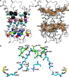
Structural localization of all the 72 additional and novel important sites. a Fifty-seven novel important sites (19 triplets) and common contact points are depicted in the BtAAC1 structure (lateral view, grey ribbon representation). The residues of triplets 80–81 (common contact points) and 22–23 are indicated as pink and tan discs, respectively; the residues of the other triplets are shown in colored beads and precisely triplet 57 (yellow), 65 (red), 35 (green), 31 and 34 (gray), 36-39 (blue), 26, 76, and 77 (cyan, located one helix turn below the common contact points), 18 and 86 (cyan, located about one helix turn above the common contact points) and 84, 85, and 88 (black). b Residues of triplets 30 and 33 (green sticks), 31, 34, 35, and 65 (cyan sticks), and 66 (yellow sticks) contribute to close the carrier on the matrix side. Ionic and H-bond interactions are indicated as dotted lines. c Other 15 novel important sites (five triplets) forming aromatic belts close to the matrix side (triplets 27, 70, and 71) and to the cytosolic side (triplets 89 and 92) are reported in stick and orange surface representations. In (a) and (c) the important sites of triplets 30, 33, 93, and 96 (m-gate and c-gate) and triplets 15, 19, 28, 66, 69, 73, and 83 (PG levels 1 and 2) are not reported
Fig. 5
Fig. 5
Inter-paralog variability of the 255 sites investigated in 53 human MCs. The inter-paralog variability of each site is expressed as the SD of the residue-specific scores of the 53 human MC amino acids. a The percentage of all important sites and remaining sites is reported in each one of ten equipotent bars covering the full range of the inter-paralog variability (SD from 0.66 to 1.96). Each bar covers a range of SD values, e.g., the first bar SD values ranging from 0.66 to 0.94. “Total” concerns the total percentage of important and remaining sites. b The percentage of sites belonging to odd and even transmembrane helices, matrix short helices and matrix loops b is reported as a function of the inter-paralog variability. The number of sites is indicated in parentheses
Fig. 6
Fig. 6
Cysteine-containing matrix short helices host the sequence motif 50-Q-Y-K-G-X-X-D-C-X-R-K-60 and previously observed acetylation of the second lysine of this motif causes local structural changes. The WebLogo representation [57] is obtained using the 15 amino acid-long matrix short helices of the human paralogs with (a) or without (b) cysteine at the sites of triplet 57. This cysteine is reported in yellow in the WebLogo representation and is underscored (C) in the sequence motif. Bits indicate amino acid conservation at each position, while the height of each symbol reflects the relative frequency of the corresponding amino acid at that position. p values of the Chi-square tests refer to the difference in the indicated amino acid frequency between the cysteine-containing and cysteine-less matrix short helices. The lower panels represent a bottom view of the ionic interaction network between K163 (the second residue of triplet 60), E153, K43, R244, and D248 in the homology structural model of the human AAC2 with (d) or without (c) the acetylation of K163. Colored sticks indicate the acetyl group (yellow), basic (cyan/green) and acid (magenta) charged residues that hold together the three repeats. Black dotted lines indicate ionic and H-bond interactions ranging between 3 and 6 Å

References

    1. Pollard KS, Hubisz MJ, Rosenbloom KR, Siepel A. Detection of non-neutral substitution rates on mammalian phylogenies. Genome Res. 2010;20:110–121. doi: 10.1101/gr.097857.109. - DOI - PMC - PubMed
    1. Veltman JA, Brunner HG. De novo mutations in human genetic disease. Nat Rev Genet. 2012;13:565–575. doi: 10.1038/nrg3241. - DOI - PubMed
    1. Lyon GJ, Wang K. Identifying disease mutations in genomic medicine settings: current challenges and how to accelerate progress. Genome Med. 2012;4:58. doi: 10.1186/gm359. - DOI - PMC - PubMed
    1. Timmermann B, Kerick M, Roehr C, Fischer A, Isau M, et al. Somatic mutation profiles of MSI and MSS colorectal cancer identified by whole exome next generation sequencing and bioinformatics analysis. PLoS One. 2010;5:e15661. doi: 10.1371/journal.pone.0015661. - DOI - PMC - PubMed
    1. Håndstad T, Rye MB, Drabløs F, Sætrom P. A ChIP-Seq benchmark shows that sequence conservation mainly improves detection of strong transcription factor binding sites. PLoS One. 2011;6:e18430. doi: 10.1371/journal.pone.0018430. - DOI - PMC - PubMed

Publication types

Substances

LinkOut - more resources