. 2013 Jun 25;108(12):2525-36.
doi: 10.1038/bjc.2013.271.

Primary breast cancer stem-like cells metastasise to bone, switch phenotype and acquire a bone tropism signature

Affiliations

Primary breast cancer stem-like cells metastasise to bone, switch phenotype and acquire a bone tropism signature

L D'Amico et al. Br J Cancer. .

Abstract

Background: Bone metastases represent a common and severe complication in breast cancer, and the involvement of cancer stem cells (CSCs) in the promotion of bone metastasis is currently under discussion. Here, we used a human-in-mice model to study bone metastasis formation due to primary breast CSCs-like colonisation.

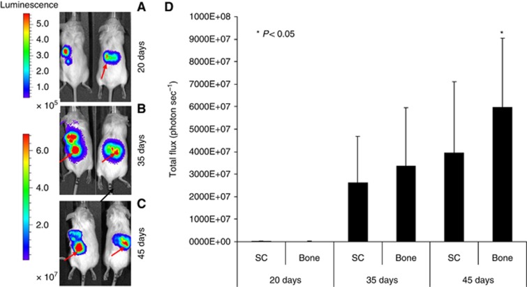
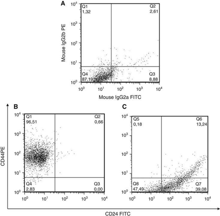
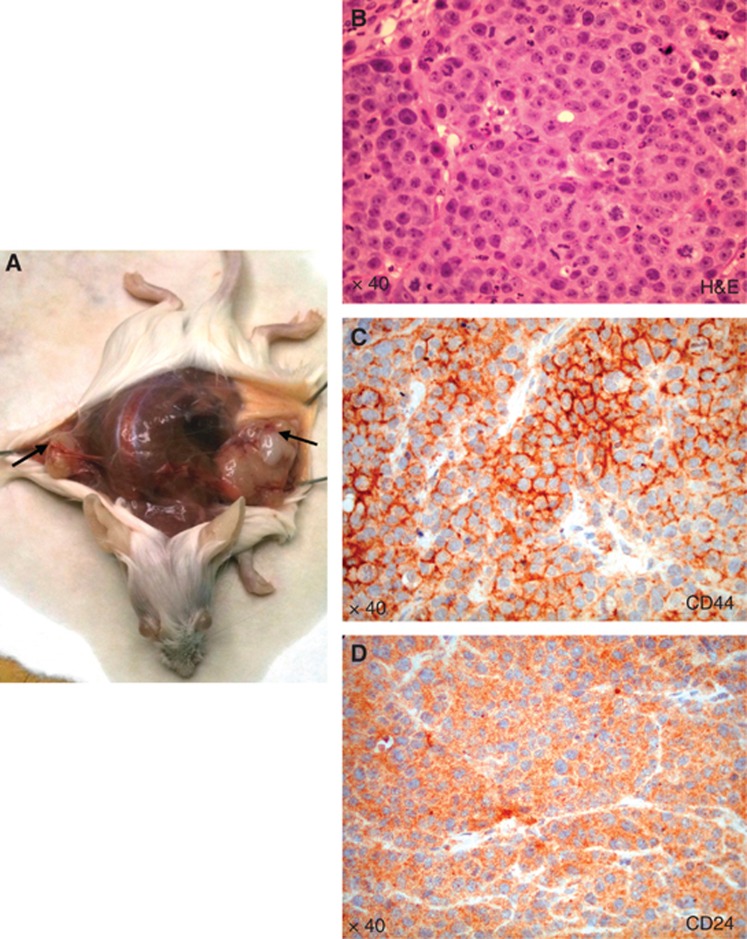
Methods: Primary CD44⁺CD24⁻ breast CSCs-like were transduced by a luciferase-lentiviral vector and injected through subcutaneous and intracardiac (IC) routes in non-obese/severe-combined immunodeficient (NOD/SCID) mice carrying subcutaneous human bone implants. The CSCs-like localisation was monitored by in vivo luciferase imaging. Bone metastatic CSCs-like were analysed through immunohistochemistry and flow cytometry, and gene expression analyses were performed by microarray techniques.

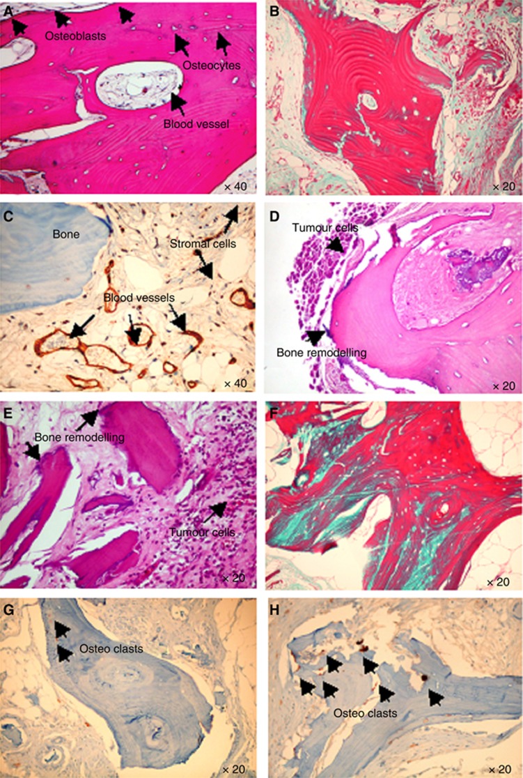
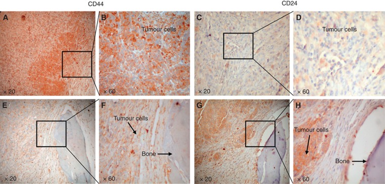
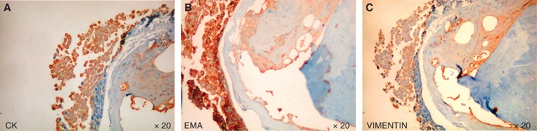
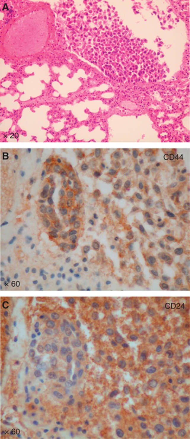
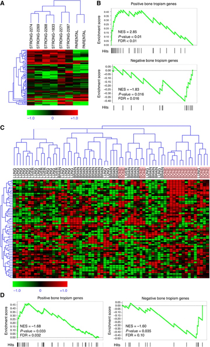
Results: Breast CSCs-like colonised the human-implanted bone, resulting in bone remodelling. Bone metastatic lesions were histologically apparent by tumour cell expression of epithelial markers and vimentin. The bone-isolated CSCs-like were CD44⁻CD24⁺ and showed tumorigenic abilities after injection in secondary mice. CD44⁻CD24⁺ CSCs-like displayed a distinct bone tropism signature that was enriched in genes that discriminate bone metastases of breast cancer from metastases at other organs.

Conclusion: Breast CSCs-like promote bone metastasis and display a CSCs-like bone tropism signature. This signature has clinical prognostic relevance, because it efficiently discriminates osteotropic breast cancers from tumour metastases at other sites.


Figures

Figure 1
Figure 1
Bioluminescence imaging (BLI). (A) Tumour growth was examined by BLI. Luc-CSCs-like were injected SC into flank of the SCID mice. Representative images of two animals after different time points (20, 35, and 45 days after SC injection) are shown. (B) At day 20, the SC tumour mass is clearly evident, whereas the bone localisation is detectable only in one mice (arrow). (C) At day 35, an increased tumour volume and invasion to human bone becomes particularly evident (arrow). (D) At 45 days, the signal derived from bone localisation of the CSCs-like is stronger in bone than that in SC tumour mass.
Figure 2
Figure 2
Histological analysis of the implanted bone. (A) H&E-stained section in control mice shows the presence of human live bone, blood vessels, newly synthesised bone with osteoblasts lining cells, as indicated by the arrows (magnification × 40). (B) Trichrome staining shows in blue the new collagen fibres in the bone: osteoid originated after the bone implanted in SC of the control mice. (C) Human endothelial cells in the blood vessels are stained for anti-human CD34, as indicated by the arrows. (D and E, respectively) Breast Luc-CSCs-like metastasise the human-implanted bone after IC and SC injection, in both cases area of pathological bone resorption are evident, as indicated by the arrows. (F) A marked neo-bone apposition is evident (osteoid is indicated by the blue staining) in bone invaded by breast CSCs-like. Osteoclasts stained for TRAP in bone of control mice (G) and mice injected with breast CSCs-like (H).
Figure 3
Figure 3
Phenotype of parental breast CSCs-like and bone-isolated CSCs-like. Representative dot plots show the isotipic control (A), the CD44+CD24 breast CSCs-like injected in mice (B) and the bone-isolated CSCs-like, which express a CD44CD24+ phenotype (C).
Figure 4
Figure 4
CD44 and CD24 expression in tumour mass and human-implanted bone. IHC staining of tumour masses shows tumour cells with a CD44+CD24 phenotype (AD). The bone-isolated CSCs-like are mainly CD44CD24+ (EH; magnification × 20 and × 60).
Figure 5
Figure 5
IHC analysis of CD44CD24+ bone-isolated CSCs-like. Cancer stem cells-like isolated from bone express CKs (A), EMA (B) and vimentin (C). (Magnification × 20).
Figure 6
Figure 6
Analysis of secondary tumour induced by CD44CD24+ bone-isolated CSCs-like. Subcutaneous injection of CD44CD24+ cells results in tumour mass formation, as indicated by the arrows in A. H&E staining of tumour mass (B), CD44 (C) and CD24 (D).
Figure 7
Figure 7
Analysis of CSCs-like lung metastases. (A) H&E-stained sections of a mouse lung show the presence of a metastatic lesion, (Magnification X 20). (B, C) IHC staining for CD44 and CD24 shows both the markers expressed by the tumor cells. (Magnification X 60).
Figure 8
Figure 8
Microarray analysis of breast CSCs-like bone tropism signature. (A) Hierarchical clustering of human breast cancer cell lines with strong bone metastatic potential derived by parental lines in Kang et al (2003) data set, using our CSCs-like bone tropism signature composed of 110 genes (119 probes). (B) Gene set enrichment analysis samples of Kang et al (2003) for enrichment of the CSCs-like bone tropism signature in genes that discriminate bone metastatics from parental lines. Upper panel: positive signature genes are significantly high in cells with strong metastatic potential. Lower panel: negative signature genes are significantly low in strong metastatic potential. (C) Hierarchical clustering of 65 breast cancer samples from metastatic lesions at different sites in a published data set, using our CSCs-like bone tropism signature. For convenience, bone metastasis samples are highlighted by light red boxes. A subset of signature genes with significantly high expression in bone metastases is evident in the top right corner. (D) The GSEA analysis on the metastasis samples for enrichment of the breast CSCs-like bone tropism signature in genes that discriminate bone metastases from lesions at other sites. Left panel: positive signature genes are significantly high in bone metastases. Right panel: negative signature genes are significantly low in bone metastases. FDR, false discovery rate; NES, normalised enrichment score.

References

    1. Abdel-Fatah TM, Powe DG, Agboola J, Adamowicz-Brice M, Blamey RW, Lopez-Garcia MA, Green AR, Reis-Filho JS, Ellis IO. The biological, clinical and prognostic implications of p53 transcriptional pathways in breast cancers. J Pathol. 2010;220 (4:419–434. - PubMed
    1. Al-Hajj M, Wicha MS, Benito-Hernandez A, Morrison SJ, Clarke MF. Prospective identification of tumorigenic breast cancer cells. Proc Natl Acad Sci USA. 2003;100 (7:3983–3988. - PMC - PubMed
    1. Beck S, Jin X, Sohn YW, Kim JK, Kim SH, Yin J, Pian X, Kim SC, Nam DH, Choi YJ, Kim H. Telomerase activity-independent function of TERT allows glioma cells to attain cancer stem cell characteristics by inducing EGFR expression. Mol Cells. 2011;31 (1:9–15. - PMC - PubMed
    1. Beider K, Abraham M, Begin M, Wald H, Weiss ID, Wald O, Pikarsky E, Abramovitch R, Zeira E, Galun E, Nagler A, Peled A. Interaction between CXCR4 and CCL20 pathways regulates tumor growth. PloS one. 2009;4 (4:e5125. - PMC - PubMed
    1. Buijs JT, van der Horst G, van den Hoogen C, Cheung H, de Rooij B, Kroon J, Petersen M, van Overveld PG, Pelger RC, van der Pluijm G. The BMP2/7 heterodimer inhibits the human breast cancer stem cell subpopulation and bone metastases formation. Oncogene. 2011;31 (17:2164–2174. - PubMed

Publication types

MeSH terms