

Bone-derived stem cells repair the heart after myocardial infarction through transdifferentiation and paracrine signaling mechanisms
- PMID: 23801066
- PMCID: PMC3822430
- DOI: 10.1161/CIRCRESAHA.113.301202
Bone-derived stem cells repair the heart after myocardial infarction through transdifferentiation and paracrine signaling mechanisms
Erratum in
-
Correction to: Bone-Derived Stem Cells Repair the Heart After Myocardial Infarction Through Transdifferentiation and Paracrine Signaling Mechanisms.Circ Res. 2021 Jan 8;128(1):e24. doi: 10.1161/RES.0000000000000451. Epub 2021 Jan 7. Circ Res. 2021. PMID: 33411635 No abstract available.
Abstract
Rationale: Autologous bone marrow-derived or cardiac-derived stem cell therapy for heart disease has demonstrated safety and efficacy in clinical trials, but functional improvements have been limited. Finding the optimal stem cell type best suited for cardiac regeneration is the key toward improving clinical outcomes.
Objective: To determine the mechanism by which novel bone-derived stem cells support the injured heart.
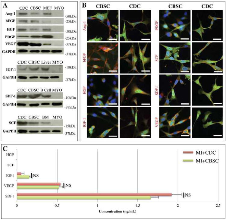
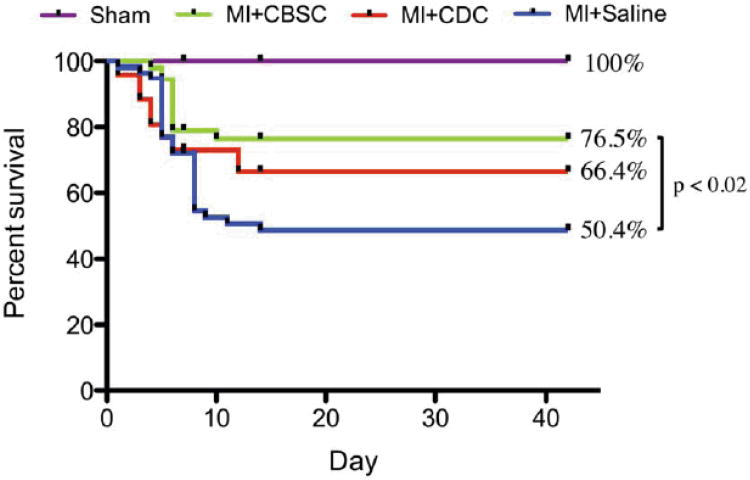
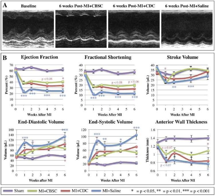
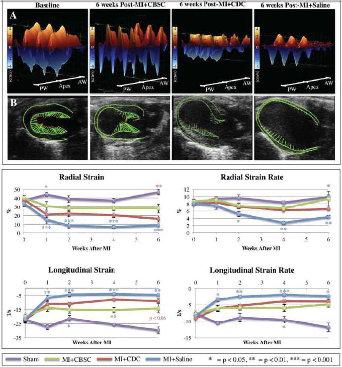
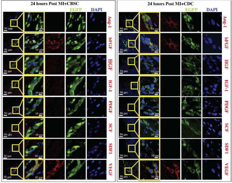
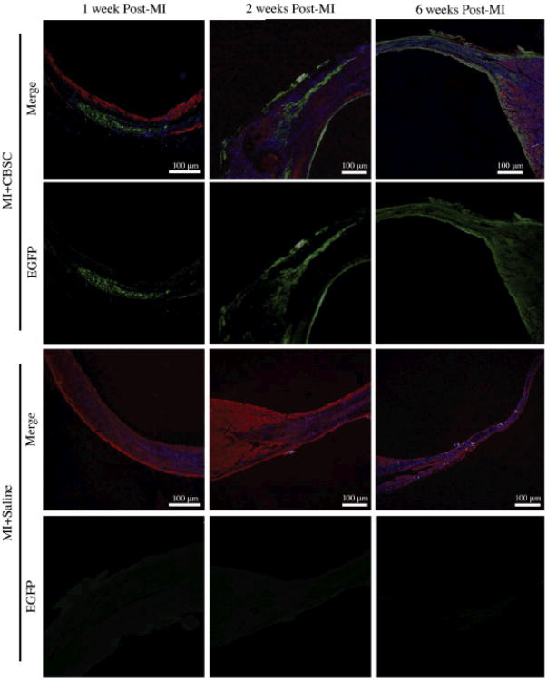
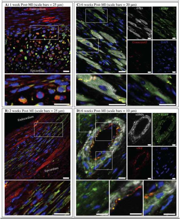
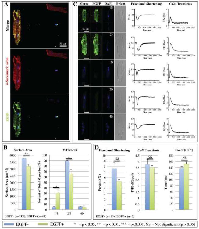
Methods and results: Cortical bone-derived stem cells (CBSCs) and cardiac-derived stem cells were isolated from enhanced green fluorescent protein (EGFP+) transgenic mice and were shown to express c-kit and Sca-1 as well as 8 paracrine factors involved in cardioprotection, angiogenesis, and stem cell function. Wild-type C57BL/6 mice underwent sham operation (n=21) or myocardial infarction with injection of CBSCs (n=67), cardiac-derived stem cells (n=36), or saline (n=60). Cardiac function was monitored using echocardiography. Only 2/8 paracrine factors were detected in EGFP+ CBSCs in vivo (basic fibroblast growth factor and vascular endothelial growth factor), and this expression was associated with increased neovascularization of the infarct border zone. CBSC therapy improved survival, cardiac function, regional strain, attenuated remodeling, and decreased infarct size relative to cardiac-derived stem cells- or saline-treated myocardial infarction controls. By 6 weeks, EGFP+ cardiomyocytes, vascular smooth muscle, and endothelial cells could be identified in CBSC-treated, but not in cardiac-derived stem cells-treated, animals. EGFP+ CBSC-derived isolated myocytes were smaller and more frequently mononucleated, but were functionally indistinguishable from EGFP- myocytes.
Conclusions: CBSCs improve survival, cardiac function, and attenuate remodeling through the following 2 mechanisms: (1) secretion of proangiogenic factors that stimulate endogenous neovascularization, and (2) differentiation into functional adult myocytes and vascular cells.
Keywords: differentiation; myocardial infarction; neovascularization; paracrine communication; stem cells.
Figures
Comment in
-
Cortical bone-derived stem cells: a novel class of cells for myocardial protection.Circ Res. 2013 Aug 16;113(5):480-3. doi: 10.1161/CIRCRESAHA.113.302083. Circ Res. 2013. PMID: 23948579 Free PMC article. No abstract available.
References
-
- Pfeffer MA, Braunwald E. Ventricular remodeling after myocardial infarction. Experimental observations and clinical implications. Circulation. 1990;81:1161–1172. - PubMed
-
- Gomez AM, Guatimosim S, Dilly KW, Vassort G, Lederer WJ. Heart failure after myocardial infarction: Altered excitation-contraction coupling. Circulation. 2001;104:688–693. - PubMed
-
- Beltrami AP, Barlucchi L, Torella D, Baker M, Limana F, Chimenti S, Kasahara H, Rota M, Musso E, Urbanek K, Leri A, Kajstura J, Nadal-Ginard B, Anversa P. Adult cardiac stem cells are multipotent and support myocardial regeneration. Cell. 2003;114:763–776. - PubMed
-
- Tang XL, Rokosh G, Sanganalmath SK, Yuan F, Sato H, Mu J, Dai S, Li C, Chen N, Peng Y, Dawn B, Hunt G, Leri A, Kajstura J, Tiwari S, Shirk G, Anversa P, Bolli R. Intracoronary administration of cardiac progenitor cells alleviates left ventricular dysfunction in rats with a 30-day-old infarction. Circulation. 2010;121:293–305. - PMC - PubMed
-
- Rota M, Padin-Iruegas ME, Misao Y, De Angelis A, Maestroni S, Ferreira-Martins J, Fiumana E, Rastaldo R, Arcarese ML, Mitchell TS, Boni A, Bolli R, Urbanek K, Hosoda T, Anversa P, Leri A, Kajstura J. Local activation or implantation of cardiac progenitor cells rescues scarred infarcted myocardium improving cardiac function. Circ Res. 2008;103:107–116. - PMC - PubMed
Publication types
MeSH terms
Substances
Grants and funding
LinkOut - more resources
Full Text Sources
Other Literature Sources
Medical
Molecular Biology Databases
Research Materials
