. 2013 Aug 9;288(32):23264-72.
doi: 10.1074/jbc.M113.491431. Epub 2013 Jun 25.

Lactosylceramide interacts with and activates cytosolic phospholipase A2α

Affiliations

Lactosylceramide interacts with and activates cytosolic phospholipase A2α

Hiroyuki Nakamura et al. J Biol Chem. .

Abstract

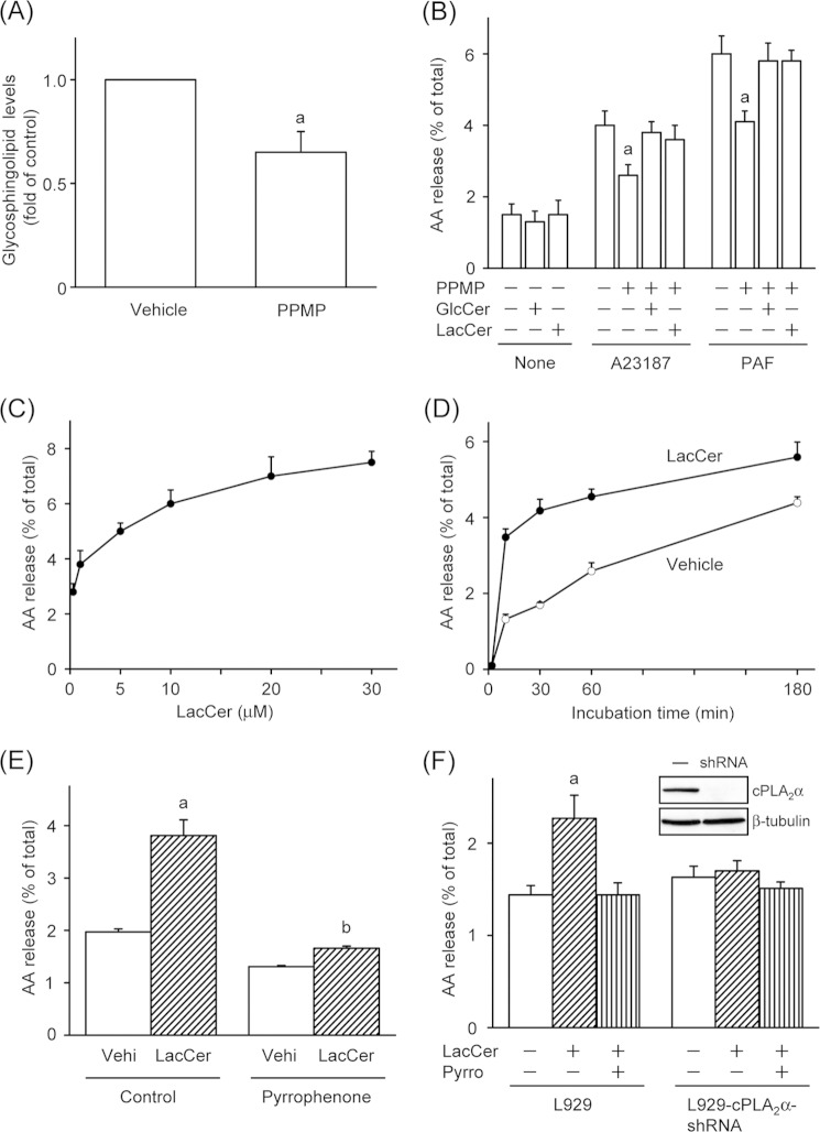
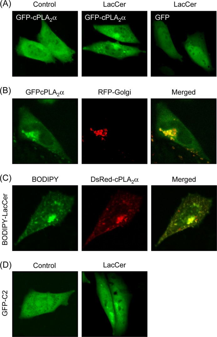
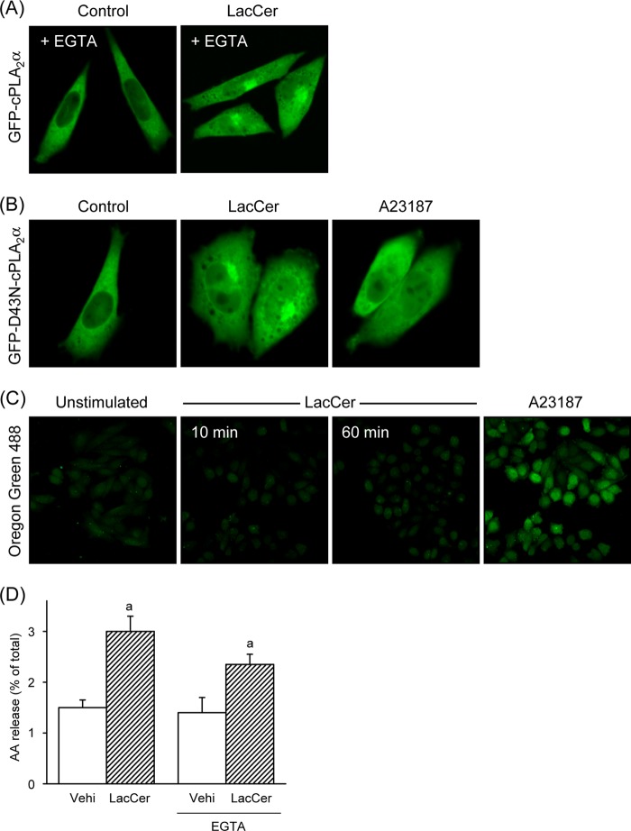
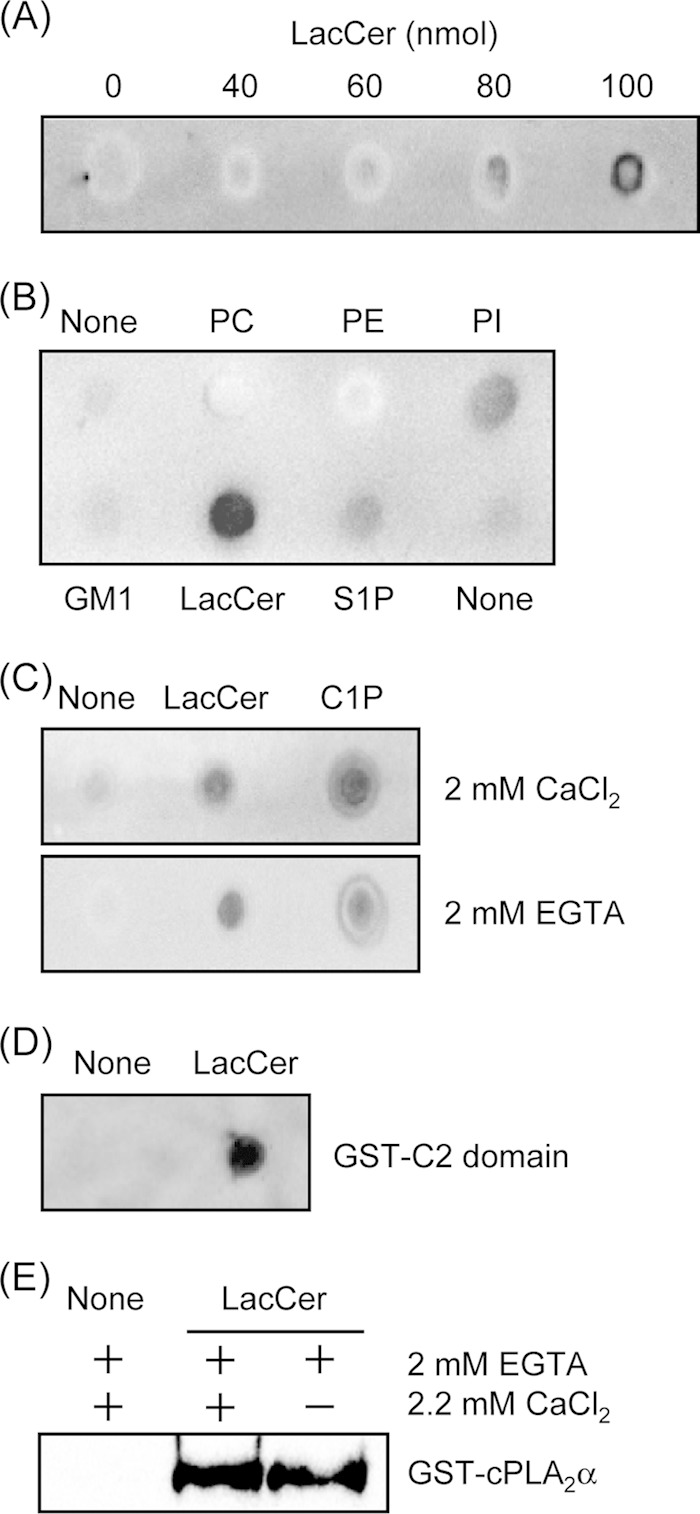
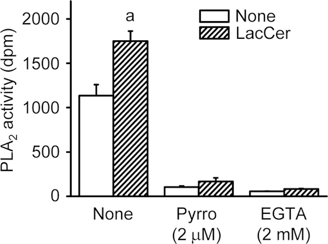
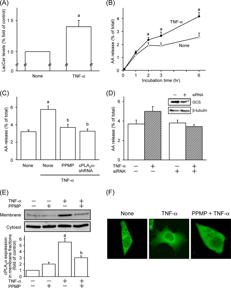
Lactosylceramide (LacCer) is a member of the glycosphingolipid family and is known to be a bioactive lipid in various cell physiological processes. However, the direct targets of LacCer and cellular events mediated by LacCer are largely unknown. In this study, we examined the effect of LacCer on the release of arachidonic acid (AA) and the activity of cytosolic phospholipase A2α (cPLA2α). In CHO-W11A cells, treatment with 1-phenyl-2-palmitoylamino-3-morpholino-1-propanol (PPMP), an inhibitor of glucosylceramide synthase, reduced the glycosphingolipid level, and the release of AA induced by A23187 or platelet-activating factor was inhibited. The addition of LacCer reversed the PPMP effect on the stimulus-induced AA release. Exogenous LacCer stimulated the release of AA, which was decreased by treatment with an inhibitor of cPLA2α or silencing of the enzyme. Treatment of CHO-W11A cells with LacCer induced the translocation of full-length cPLA2α and its C2 domain from the cytosol to the Golgi apparatus. LacCer also induced the translocation of the D43N mutant of cPLA2α. Treatment of L929 cells with TNF-α induced LacCer generation and mediated the translocation of cPLA2α and AA release, which was attenuated by treatment with PPMP. In vitro studies were then conducted to test whether LacCer interacts directly with cPLA2α. Phosphatidylcholine vesicles containing LacCer increased cPLA2α activity. LacCer bound to cPLA2α and its C2 domain in a Ca(2+)-independent manner. Thus, we propose that LacCer is a direct activator of cPLA2α.

Keywords: Arachidonic acid; Glycosphingolipid; Phospholipase A; Sphingolipid; Tumor Necrosis Factor (TNF).


Figures

FIGURE 1.
FIGURE 1.
LacCer induces cPLA2α-dependent AA release from cells. A, CHO-W11A cells were cultured for 48 h in culture medium supplemented with vehicle or 1 μm PPMP. After the cells were washed, glycosphingolipids were extracted and spotted onto a TLC plate. B, CHO-W11A cells were cultured for 30 h in culture medium supplemented with or without 1 μm PPMP, 10 μm GlcCer, and/or 10 μm LacCer. The cells were then labeled by incubation for 18 h in 0.1% BSA- and [3H]AA-containing medium supplemented with the same reagents. The labeled cells were washed and stimulated with 1 μm A23187 or 100 nm PAF for 30 min at 37 °C. a, p < 0.05, significantly different from the control without PPMP. C, [3H]AA-labeled cells were stimulated with the indicated concentrations of LacCer for 30 min at 37 °C. D, [3H]AA-labeled cells were stimulated with vehicle or 30 μm LacCer for the indicated time periods at 37 °C. ○, vehicle; ●, LacCer. E, the labeled cells were pretreated with vehicle (Control) or 2 μm pyrrophenone for 30 min prior to stimulation with vehicle (Vehi) or 30 μm LacCer for 30 min at 37 °C. a, p < 0.05, significantly different from the control without LacCer; b, p < 0.05, significantly different from the control without pyrrophenone. F, [3H]AA-labeled L929 and L929-cPLA2α-shRNA cells were pretreated with 2 μm pyrrophenone (Pyrro) for 30 min and then stimulated with 30 μm LacCer for 30 min at 37 °C. The expression levels of cPLA2α in both cells are shown. a, p < 0.05, significantly different from the control without LacCer. The data shown are the means ± S.E. for three experiments.
FIGURE 2.
FIGURE 2.
LacCer induces cPLA2α translocation in cells. A, CHO-W11A cells transiently transfected with expression vectors for GFP-cPLA2α and GFP were treated with vehicle (Control) or 30 μm LacCer for 1 h. B, CHO-W11A cells transiently transfected with expression vectors for GFP-cPLA2α and red fluorescent protein (RFP)-Golgi were treated with 30 μm LacCer. C, DsRed-cPLA2α-expressing CHO-W11A cells were treated with 100 nm BODIPY-C5-LacCer for 1 h. D, GFP-C2-expressing CHO-W11A cells were treated with 30 μm LacCer for 1 h. In A–D, data are representative of three independent experiments.
FIGURE 3.
FIGURE 3.
LacCer activates cPLA2α without intracellular Ca2+ mobilization. A, CHO-W11A cells transiently transfected with expression vector for GFP-cPLA2α were treated with vehicle (Control) or 30 μm LacCer for 1 h in the presence of 2 mm EGTA. B, GFP-D43N-cPLA2α-expressing CHO-W11A cells were treated with 30 μm LacCer or 5 μm A23187 for 1 h. C, CHO-W11A cells labeled with Oregon Green 488 were washed and then treated with 30 μm LacCer for 10 or 60 min or with 5 μm A23187 for 5 min. In A–C, data are representative of three independent experiments. D, [3H]AA-labeled CHO-W11A cells were stimulated with vehicle (Vehi) or 30 μm LacCer for 30 min at 37 °C in the presence or absence of 2 mm EGTA. a, p < 0.05, significantly different from the control without LacCer.
FIGURE 4.
FIGURE 4.
cPLA2α binds to LacCer in a calcium-independent manner. A, the binding of cPLA2α to LacCer was examined using the lipid-protein overlay assay as described under “Experimental Procedures.” The indicated amounts of LacCer were spotted onto a Hybond C membrane. The membrane was exposed to purified GST-cPLA2α overnight at 4 °C. B, the lipid-protein overlay assay was repeated with 100 nmol of lipids and GST-cPLA2α. PC, 1-palmitoyl-2-oleoylphosphatidylcholine; PE, 1-palmitoyl-2-oleoylphosphatidylethanolamine; PI, 1-palmitoyl-2-oleoylphosphatidylinositol; S1P, sphingosine 1-phosphate. C, the lipid-protein overlay assay was repeated with 100 nmol of LacCer, 20 nmol of C1P, and GST-cPLA2α with or without 2 mm EGTA. D, the membrane spotted with 100 nmol of LacCer was exposed to the GST-tagged C2 domain of cPLA2α overnight at 4 °C. E, the large multilamellar vesicle binding assay was performed as described under “Experimental Procedures.” In A–E, data are representative of three independent experiments.
FIGURE 5.
FIGURE 5.
LacCer activates cPLA2α in vitro. PLA2 activity in the cytosolic fraction from HEK293T cells expressing human cPLA2α was measured as described under “Experimental Procedures.” PLA2 activities toward PAPC and 95:5 PAPC/LacCer-mixed vesicles were measured in reaction buffer with or without 2 μm pyrrophenone (Pyrro) or 2 mm EGTA. The data shown are the means ± S.E. for three experiments. a, p < 0.05, significantly different from the control without LacCer.
FIGURE 6.
FIGURE 6.
TNF-α induces cPLA2α-dependent AA release via LacCer formation in L929 cells. A, L929 cells labeled with [14C]serine for 18 h were stimulated with or without 10 nm TNF-α for 2 h. After the cells were washed, lipids were extracted and separated by TLC as described under “Experimental Procedures.” B, [3H]AA-labeled L929 cells were stimulated with 10 nm TNF-α for the indicated time periods at 37 °C. ○, none; ●, TNF-α. C, L929 cells were pretreated with vehicle or 3 μm PPMP for 30 min, and then the cells and L929-cPLA2α-shRNA cells were stimulated with 10 nm TNF-α for 6 h at 37 °C. D, down-regulation of GlcCer synthase were performed as described under “Experimental Procedures.” L929 cells transfected with or without GlcCer synthase siRNA were stimulated with 10 nm TNF-α for 6 h at 37 °C. The expression levels of GlcCer synthase (GCS) in cells are shown. E, L929 cells were pretreated with vehicle or 3 μm PPMP for 30 min and then stimulated with 10 nm TNF-α for 2 h at 37 °C. The cytosolic and membrane fractions were prepared and subjected to immunoblot analysis. Upper panels, immunoblotting with antibodies against cPLA2α. Lower panel, the histogram represents the expression levels of cPLA2α in membrane fractions quantified using ImageJ. The data shown are the means ± S.E. for three experiments. a, p < 0.05, significantly different from the control without TNF-α; b, p < 0.05, significantly different from the control without PPMP or cPLA2α knockdown. F, L929 cells transiently transfected with expression vector for GFP-cPLA2α were treated with or without TNF-α for 2 h. PPMP were pretreated for 30 min before stimulation with TNF-α.

Similar articles

Cited by

References

    1. Murakami M., Taketomi Y., Miki Y., Sato H., Hirabayashi T., Yamamoto K. (2011) Recent progress in phospholipase A2 research: from cells to animals to humans. Prog. Lipid Res. 50, 152–192 - PubMed
    1. Hirabayashi T., Murayama T., Shimizu T. (2004) Regulatory mechanism and physiological role of cytosolic phospholipase A2. Biol. Pharm. Bull. 27, 1168–1173 - PubMed
    1. Evans J. H., Spencer D. M., Zweifach A., Leslie C. C. (2001) Intracellular calcium signals regulating cytosolic phospholipase A2 translocation to internal membranes. J. Biol. Chem. 276, 30150–30160 - PubMed
    1. Tucker D. E., Ghosh M., Ghomashchi F., Loper R., Suram S., John B. S., Girotti M., Bollinger J. G., Gelb M. H., Leslie C. C. (2009) Role of phosphorylation and basic residues in the catalytic domain of cytosolic phospholipase A2α in regulating interfacial kinetics and binding and cellular function. J. Biol. Chem. 284, 9596–9611 - PMC - PubMed
    1. Subramanian P., Vora M., Gentile L. B., Stahelin R. V., Chalfant C. E. (2007) Anionic lipids activate group IVA cytosolic phospholipase A2 via distinct and separate mechanisms. J. Lipid Res. 48, 2701–2708 - PubMed

Publication types

MeSH terms

LinkOut - more resources