. 2012 Jan-Feb;1(1):3-15.
doi: 10.1002/wdev.2. Epub 2011 Nov 17.

Morphogen gradients in development: from form to function

Affiliations

Morphogen gradients in development: from form to function

Jan L Christian. Wiley Interdiscip Rev Dev Biol. 2012 Jan-Feb.

Abstract

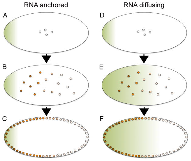
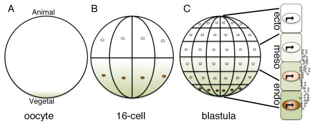
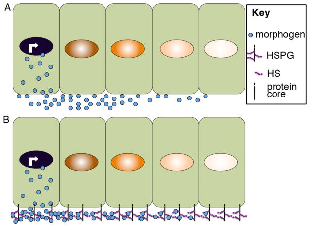
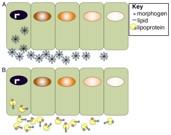
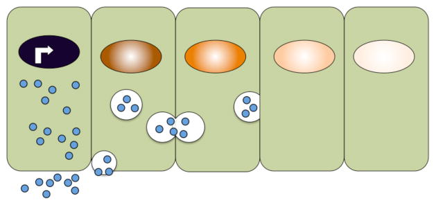
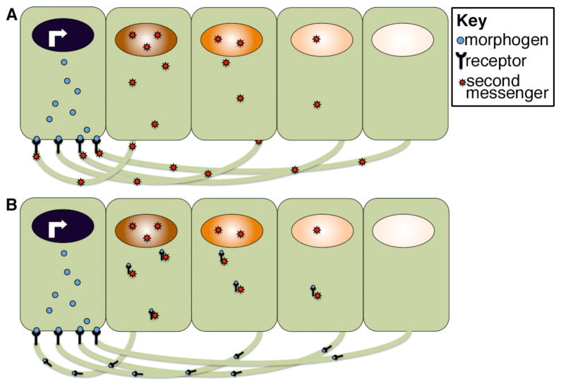
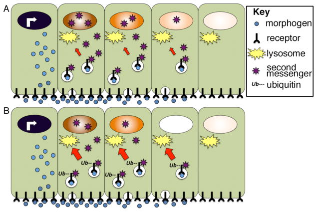
Morphogens are substances that establish a graded distribution and elicit distinct cellular responses in a dose-dependent manner. They function to provide individual cells within a field with positional information, which is interpreted to give rise to spatial patterns. Morphogens can consist of intracellular factors that set up a concentration gradient by diffusion in the cytoplasm. More commonly, morphogens comprise secreted proteins that form an extracellular gradient across a field of cells. Experimental studies and computational analyses have provided support for a number of diverse strategies by which extracellular morphogen gradients are formed. These include free diffusion in the extracellular space, restricted diffusion aided by interactions with heparan sulfate proteoglycans, transport on lipid-containing carriers or transport aided by soluble binding partners. More specialized modes of transport have also been postulated such as transcytosis, in which repeated rounds of secretion, endocytosis, and intracellular trafficking move morphogens through cells rather than around them, or cytonemes, which consist of filopodial extensions from signal-receiving cells that are hypothesized to reach out to morphogen-sending cells. Once the gradient has formed, cells must distinguish small differences in morphogen concentration and store this information even after the gradient has dissipated. This is often achieved by translating ligand concentration into a proportional increase in numbers of activated cell surface receptors that are internalized and continue to signal from endosomal compartments. Ultimately, this leads to activation of one or a few transcription factors that transduce this information into qualitatively distinct gene responses inside the nucleus.


Figures

Figure 1
Figure 1. Illustration of the Bicoid gradient in the early syncitial Drosophila embryo
(A–F) Following fertilization of the Drosophila egg, nuclei divide in the absence of cell division and remain positioned near the center of the embryo for the first 8 cell cycles (A–B, D–E) but then migrate to the periphery (C,F). bicoid RNA (green shading) is deposited at the anterior pole of the egg (A,D) and a nuclear Bicoid protein gradient (orange shading) forms either by translation of the localized RNA followed by protein diffusion away from the anterior pole (AC) or by diffusion of RNA away from the anterior pole followed by local translation of the graded RNA (D–F). Anterior is to the left in all panels.
Figure 2
Figure 2. The VegT-nodal morphogen gradient in the early Xenopus embryo
(A–C) vegT RNA (green) is anchored to the cortical cytoskeleton at the vegetal pole of the oocyte (A) but is released upon oocyte maturation and diffuses toward the animal pole during early cleavage stages (B). Local translation of vegT RNA generates a gradient of VegT protein (orange) that is restricted to nuclei of prospective endodermal cells located near the vegetal pole (B,C). VegT activates transcription of target genes such as nodal and other TGF-β family members, which encode secreted morphogens (C, small circles). Nodal is proposed to specify endodermal (endo) and mesodermal (meso) fate at high and moderate doses, respectively. Cells that are not exposed to nodal adopt an ectodermal (ecto) fate.
Figure 3
Figure 3. Extracellular movement of morphogens assisted by interactions with HSPGs
Illustration of morphogen movement through the extracellular space by free diffusion (A) or facilitated by interactions with HSPGs (B). Illustration is schematic and not meant to imply that morphogen movement is restricted to one side of the cell.
Figure 4
Figure 4. Proposed modes of extracellular transport of lipid-linked morphogens
Lipid-linked morphogens may form micellar-like aggregates in which lipids are positioned on the inside and are surrounded by hydrophobic residues (A) or they may be transported on lipoprotein complexes (B). Illustration is schematic and not meant to imply that morphogen movement is restricted to one side of the cell.
Figure 5
Figure 5. Extracellular transport of Dpp facilitated by Sog
Sog competes with receptors for binding to Dpp and restricts its ability to signal to ventral cells while facillitating its diffusion toward the dorsal side of the embryo. Tollid cleaves Sog, releasing Dpp and enabling it to bind and activate receptors. Illustration is schematic and not meant to imply that morphogen movement is restricted to one side of the cell.
Figure 6
Figure 6. Model for morphogen transport by transcytosis
Schematic illustration of a model in which morphogens are actively moved between cells by repeated rounds of secretion, endocytosis and intracellular transport in vesicles.
Figure 7
Figure 7. Model for cytoneme function in morphogen gradient formation
Morphogen receiving cells extend long actin-based filopodia toward morphogen secreting cells. Receptors are proposed to bind the morphogen at points of contact and to either activate second messengers that then traffic back to the body of the target cell (A) or to directly traffic the bound morphogen to receiving cells (B). Illustration is schematic and not meant to imply that morphogen movement is restricted to one side of the cell.
Figure 8
Figure 8. Lysosomal trafficking of activated receptors can shape the morphogen activity gradient
(A) Following ligand binding, receptor-ligand complexes are internalized and continue to activate second messengers from endosomal compartments. Receptors can be trafficked back to the membrane, or they can be trafficked to the lysosome (red arrow) where they are degraded. (B) Cellular influences that accelerate receptor ubiquitinylation lead to a corresponding increase in the rate of lysosomal trafficking (red arrow) and degradation. This shortens the duration of intracellular signaling, and hence the strength of the nuclear response to an identical extracellular morphogen gradient. Illustration is schematic and not meant to imply that morphogen movement is restricted to one side of the cell.

References

    1. Turing AM. The chemical basis of morphogenesis. Philos Trans R Soc Lond B Biol Sci. 1952;237:37–72. - PMC - PubMed
    1. Wolpert L. Positional information and the spatial pattern of cellular differentiation. J Theor Biol. 1969;25:1–47. - PubMed
    1. Wolpert L. Positional information and patterning revisited. J Theor Biol. 2011;269:359–365. - PubMed
    1. Gao B, Song H, Bishop K, Elliot G, Garrett L, English MA, Andre P, Robinson J, Sood R, Minami Y, et al. Wnt signaling gradients establish planar cell polarity by inducing Vangl2 phosphorylation through Ror2. Dev Cell. 2011;20:163–176. - PMC - PubMed
    1. Schwank G, Basler K. Regulation of organ growth by morphogen gradients. Cold Spring Harb Perspect Biol. 2010;2:a001669. - PMC - PubMed

Further Reading/Resources

    1. Gilbert . Developmental Biology. 9. 2010.
    1. The interactive fly. http://www.sdbonline.org/fly/aimain/1aahome.htm.
    1. Port F, Basler K. Wnt trafficking: new insights into Wnt maturation, secretion and spreading. Traffic. 2010;11:1265–1271. - PubMed
    1. Gallet A. Hedgehog morphogen: from secretion to reception. Trends Cell Biol. 2011;21:238–246. - PubMed
    1. Schier AF. Nodal morphogens. Cold Spring Harb Perspect Biol. 2009;1:a003459. - PMC - PubMed

MeSH terms

LinkOut - more resources