Comparative Study
. 2012 Jan-Feb;1(1):16-39.
doi: 10.1002/wdev.3. Epub 2011 Nov 17.

Comparisons of the embryonic development of Drosophila, Nasonia, and Tribolium

Affiliations
Comparative Study

Comparisons of the embryonic development of Drosophila, Nasonia, and Tribolium

Ezzat El-Sherif et al. Wiley Interdiscip Rev Dev Biol. 2012 Jan-Feb.

Abstract

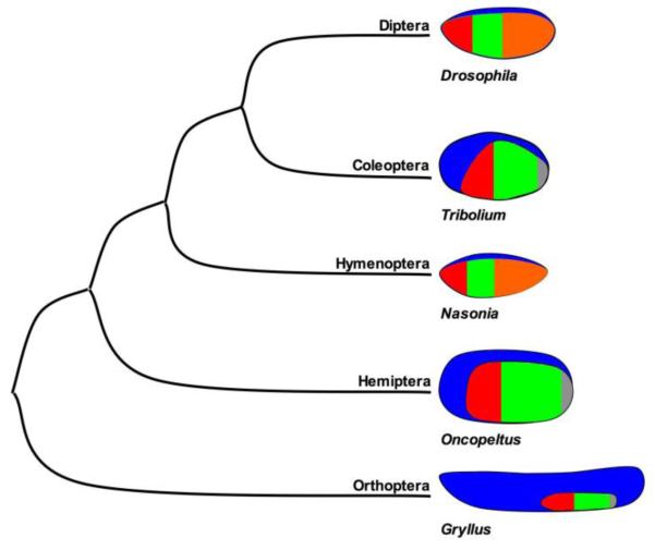
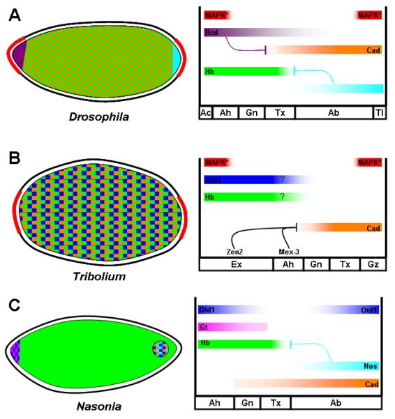
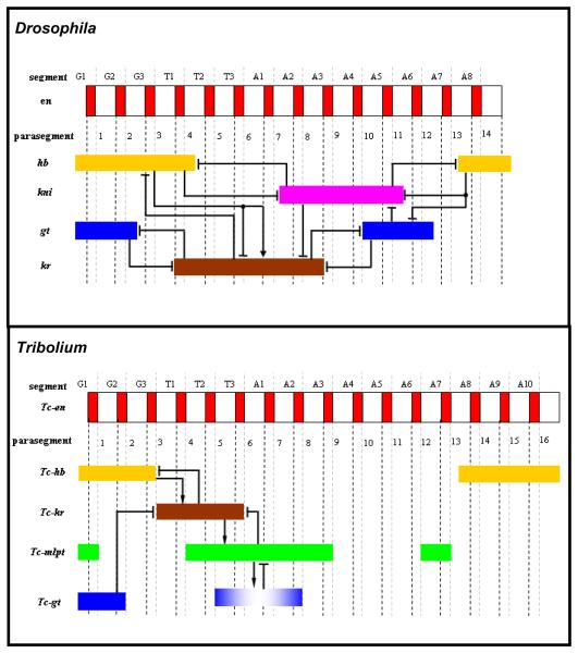
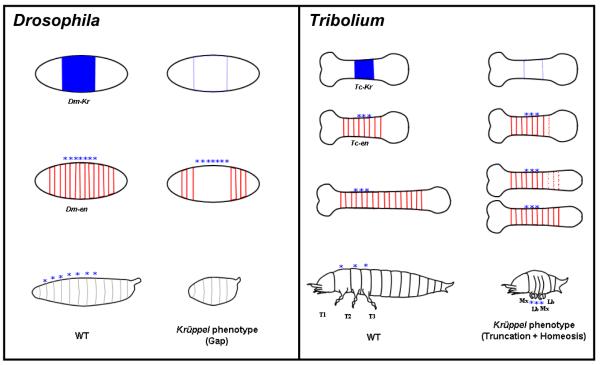
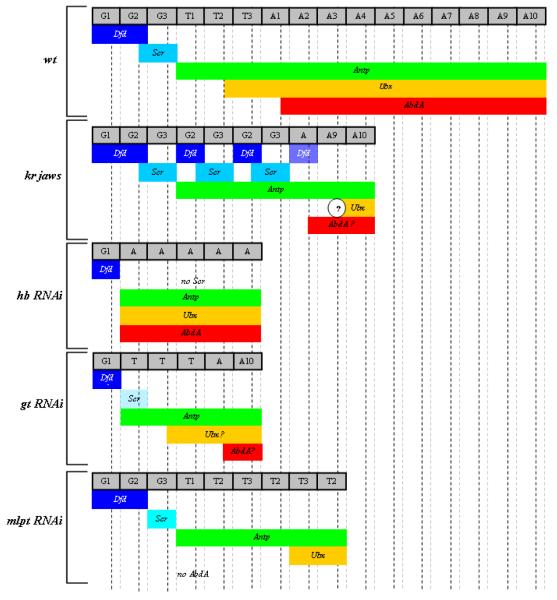
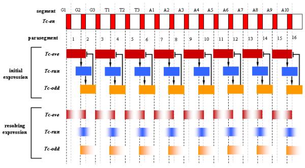
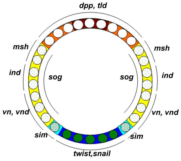
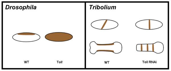
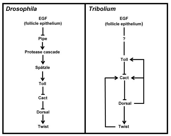
Studying the embryogenesis of diverse insect species is crucial to understanding insect evolution. Here, we review current advances in understanding the development of two emerging model organisms: the wasp Nasonia vitripennis and the beetle Tribolium castaneum in comparison with the well-studied fruit fly Drosophila melanogaster. Although Nasonia represents the most basally branching order of holometabolous insects, it employs a derived long germband mode of embryogenesis, more like that of Drosophila, whereas Tribolium undergoes an intermediate germband mode of embryogenesis, which is more similar to the ancestral mechanism. Comparing the embryonic development and genetic regulation of early patterning events in these three insects has given invaluable insights into insect evolution. The similar mode of embryogenesis of Drosophila and Nasonia is reflected in their reliance on maternal morphogenetic gradients. However, they employ different genes as maternal factors, reflecting the evolutionary distance separating them. Tribolium, on the other hand, relies heavily on self-regulatory mechanisms other than maternal cues, reflecting its sequential nature of segmentation and the need for reiterated patterning.


Figures

Figure 1
Figure 1. Phylogenetic relationships of selected insect models, and schematic representations of their early embryonic fate maps
Blue = extraembryonic tissue, Red = segments of the head, Green = thoracic segments, Orange = abdominal segments, and Gray = growth zone primordium.
Figure 2
Figure 2. Maternal provision of AP patterning information in Drosophila, Tribolium, and Nasonia
Left: Schematic representation of Drosophila (A), Tribolium (B), and Nasonia (C) embryos showing distribution of maternally provided mRNAs. Right: representation of resulting protein gradients. Red curves in left panel on A and B represent the distribution of tsl expression, and correspond with the red bars at right representing the resulting activation of MAP kinase in the embryo. Question marks for Otd and Hb in panel B indicate their now doubted role in providing positional information to the early embryo. Ah=anterior head, Gn=gnathal, Tx=thorax, Ab=abdomen, Tl=telson, Gz=growth zone.
Figure 3
Figure 3. Comparison of gap gene expression patterns and their regulatory relationships in Drosophila and Tribolium
Top panel: Late blastodermal expression patterns of gap genes (non-terminal, non-head) in Drosophila and their regulatory relationships. Bottom panel: post-blastodermal expression patterns of gap genes (non-terminal, non-head) in Tribolium after they emanate from the growth zone and before they fade, and a parsimonious interpretation of their regulatory relationships. An arrow represents positive regulation and a blunt line represents negative regulation. G= gnathal segment, T= thoracic segment, A= abdominal segment.
Figure 4
Figure 4. Comparison of gap genes phenotypes in Drosophila and Tribolium
Expression of Krüppel (in blue) and engrailed (in red) and the resulting larval cuticles (bottom of each panel) in WT (left of each panels) and Krüppel mutants (right of each panel). Left panel (Drosophila): The strongest Krüppel phenotype in Drosophila results in loss of seven segments (marked by stars) within the expression domain of the gene Krüppel, which is a typical “gap” phenotype. Right panel (Tribolium): In the Krüppel mutant (jaws), the segments within the Tc-Krüppel expression domain (marked by stars) form properly, and only a few more posterior segments develop, which are disorganized at first but later regulated to form intact segments. In addition to this “truncation” phenotype, a homeotic transformation is observed in the larval cuticle, in which the three thoracic segments and the first abdominal segment are transformed to gnathal identity (maxillary-labial-maxillary-labial). This phenotype is best described as “truncation plus homeosis”, rather than “gap”. The yet unexplained “truncation after expression domain” of Tribolium gap genes in contrast to “gap within expression domain” of Drosophila is proved valuable here to show clearly the homeotic effect of gap genes phenotypes in Tribolium, whereas it is precluded by the loss of segments in Drosophila (except for some hypomorphic mutants).
Figure 5
Figure 5. Hox gene expression in wt and gap gene RNAi and mutant embryos
The identity of segments that are formed is described in the gray boxes at the top of each panel. Diluted color in an expression domain indicates reduced expression. A question mark inside an expression domain indicates that this expression domain is not reported in literature. A question mark in a circle at the border of an expression domain indicates that the exact location of this border is not reported precisely in literature. G= gnathal segments, T= thoracic segments, A= abdominal segments.
Figure 6
Figure 6. Initial and resolving expressions of primary pair-rule genes in Tribolium
A negative feedback loop between the three primary pair-rule genes Tc-eve, Tc-run, and Tc-odd are responsible for their initial double-segmental periodic expression. Later, they resolve into segmental periodic expression through a yet to be identified genetic mechanism. This later segmental primary pair-rule expression regulates downstream genes to ultimately position segment polarity genes. An arrow represents positive regulation and a blunt line represents negative regulation. G= gnathal segment, T= thoracic segment, A= abdominal segment. It is wothnoting that these regulatory relationships were verified for the anteriormost segments. Further expriments are needed to verify it for the following segments (using embryonic RNAi).
Figure 7
Figure 7. DV fate map and expression domains of genes involved in DV patterning in Drosophila
Different concentrations of nuclear Dorsal drive different sets of downstream genes and ultimately (with the help BMP and EGF signaling) define DV fates. Blue: mesoderm, cyan: mesectoderm, yellow: neurogenic ectoderm, orange: dorsal ectoderm, red: amnioserosa, opaque green circles: high nuclear Dorsal concentration, heavily dotted green circles: intermediate nuclear Dorsal concentration, lightly dotted green circles: low nuclear Dorsal concentration, white nuclei: no nuclear Dorsal.
Figure 8
Figure 8. Comparison between Toll phenotype in Drosophila and Tribolium
Left panel (Drosophila): dpp (brown) is expressed dorsally in WT (left) and along the entire DV axis in a Toll mutant (right). Right panel (Tribolium): Early blastodermal Tc-dpp is expressed as an oblique stripe in WT (left upper embryo), and loses this obliqueness in Toll RNAi (right upper embryo). In Tribolium, the initial orientation of balstodermal Tc-dpp dictates the orientation of later reiterated germband expression of Tc-dpp. Consequently, Tc-dpp reiterates only twice in the DV direction in WT probably due to space constraints (left lower embryo), and many times in the AP direction in Toll RNAi (right lower embryo).
Figure 9
Figure 9. Regulatory relationships between major components in DV axis patterning
Left panel (Drosophila): the DV axis patterning network in Drosophila is mostly a linear cascade with the exception of multiple positive and negative feedback loops within the protease cascade components. Right panel (Tribolium): nothing is known about the involvement of a protease cascade in Tribolium similar to that involved in Drosophila. However, Tribolium posses multiple feedback loops at levels more downstream.
Figure 10
Figure 10. Germ cell formation in Drosophila, Nasonia, and Tribolium
Red=osk mRNA, magenta=Tc-vasa mRNA, blue=nuclei. A1-C1: In the early cleavage stages of embryogenesis, osk mRNA is localized in the posterior pole plasm of Drosophila (A1) and oosome of Nasonia (B1), while Tc-vasa is ubiquitous in Tribolium (C1). A2-C2: As the syncytial blastoderm forms, posterior nuclei interact with the pole plasm (A2), or the oosome (B2) in fly and wasp, respectively, while nuclei enter an apparently homogenous environment in Tribolium (C2). A3-C3: Individual nuclei that enter the posterior pole plasm bud singly, forming the pole cells in Drosophila (A3). In contrast, the oosome along with multiple nuclei bud simultaneously from the posterior in Nasonia (B3). In the later blastoderm stages of Tribolium, Tc-vasa mRNA is cleared from the embryo, save for the most posterior pole. A4-C4: Production of pole cells is completed, and osk mRNA begins to be degraded in the cellular blastoderm stage in both Drosophila (A4) and Nasonia (B4). Posterior cells that retained Tc-vasa expression delaminate from the blastoderm into the interior of the embryo (C4) and will become the primordial germ cells of the beetle.
Figure 11
Figure 11. Mechanisms of gastrulation in Drosophila, Tribolium, and Nasonia
A-A”: Gastrulation by furrow formation employed by Drosophila, and Tribolium in the anterior segments. B-B”: Gastrulation mode found in the segments deriving from the growth zone of Tribolium. A multilayered mass of cells loses its epithelial character and is internalized by the migration of the lateral ectodermal cells. C-C”: Hymenopteran mode of gastrulation, where the mesoderm forms a stiff plate, the ectoderm breaks contact with mesoderm and migrates ventrally until the free edges meet and fuse at the ventral midline. Blue= lateral ectoderm, Green=mesectoderm, Red= mesoderm precursors expressing twist, Pink= mesoderm precursors not expressing twist. Other cell fates have been omitted for clarity.

References

    1. Davis GK, Patel NH. Short, long, and beyond: Molecular and embryological approaches to insect segmentation. Annual Review of Entomology. 2002;47:669–699. - PubMed
    1. Tautz D, Friedrich M, Schröder R. Insect embryogenesis - what is ancestral and what is derived? Development. 1994;(Supplement):193–199.
    1. Werren JH, Richards S, Desjardins CA, Niehuis O, Gadau J, Colbourne JK, Beukeboom LW, Desplan C, Elsik CG, Grimmelikhuijzen CJP, et al. Functional and Evolutionary Insights from the Genomes of Three Parasitoid Nasonia Species. Science. 2010;327:343–348. - PMC - PubMed
    1. Richards S, Gibbs RA, Weinstock GM, Brown SJ, Denell R, Beeman RW, Gibbs R, Bucher G, Friedrich M, Grimmelikhuijzen CJP, et al. The genome of the model beetle and pest Tribolium castaneum. Nature. 2008;452:949–955. - PubMed
    1. Bucher G, Scholten J, Klingler M. Parental RNAi in Tribolium (Coleoptera) Curr Biol. 2002;12:R85–86. - PubMed

Publication types

MeSH terms

Substances

LinkOut - more resources