

Genotypes and phenotypes of 162 families with a glomulin mutation
- PMID: 23801931
- PMCID: PMC3666456
- DOI: 10.1159/000348675
Genotypes and phenotypes of 162 families with a glomulin mutation
Abstract
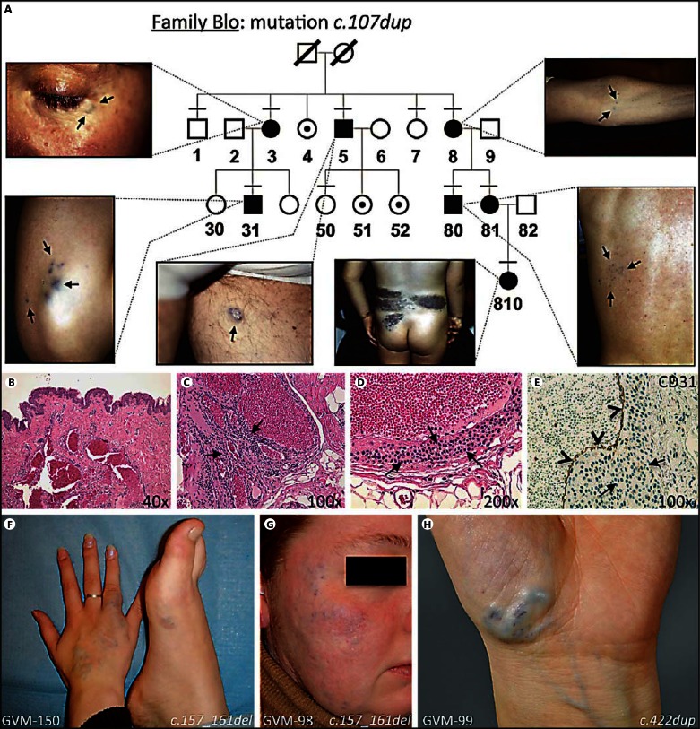
A decade ago, we identified a novel gene, glomulin (GLMN) in which mutations cause glomuvenous malformations (GVMs). GVMs are bluish-purple cutaneous vascular lesions with characteristic glomus cells in the walls of distended venous channels. The discovery of the genetic basis for GVMs allowed the definition of clinical features to distinguish GVMs from other venous anomalies. The variation in phenotype was also highlighted: from a single punctate blue dot to a large plaque-like lesion. In this study, we screened GLMN in a large cohort of patients to broaden the spectrum of mutations, define their frequency and search for possible genotype-phenotype correlations. Taking into account 6 families published by others, a mutation in GLMN has been found in 162 families. This represents 40 different mutations; the most frequent one being present in almost 45% of them. Expressivity varies largely, without a genotype/phenotype relationship. Among 381 individuals with a mutation, we discovered 37 unaffected carriers, implying a penetrance of 90%. As nonpenetrant individuals may transmit the disease to their descendants, knowledge on the mutational status is needed for appropriate genetic counseling.
Keywords: Anomaly; Gene; Glomulin; Glomuvenous malformation; Vascular.
Figures

References
-
- Al Dhaybi R, Powell J, McCuaig C, Kokta V. Differentiation of vascular tumors from vascular malformations by expression of Wilms tumor 1 gene: evaluation of 126 cases. J Am Acad Dermatol. 2010;63:1052–1057. - PubMed
-
- Blume-Peytavi U, Adler YD, Geilen CC, Ahmad W, Christiano A, et al. Multiple familial cutaneous glomangioma: a pedigree of 4 generations and critical analysis of histologic and genetic differences of glomus tumors. J Am Acad Dermatol. 2000;42:633–639. - PubMed
-
- Boon LM, Mulliken JB, Enjolras O, Vikkula M. Glomuvenous malformation (glomangioma) and venous malformation: distinct clinicopathologic and genetic entities. Arch Dermatol. 2004;140:971–976. - PubMed
Grants and funding
LinkOut - more resources
Full Text Sources
Other Literature Sources
Research Materials
