. 2013 Jun 20:4:76.
doi: 10.3389/fneur.2013.00076. eCollection 2013.

Trinucleotide repeats: a structural perspective

Affiliations

Trinucleotide repeats: a structural perspective

Bruno Almeida et al. Front Neurol. .

Abstract

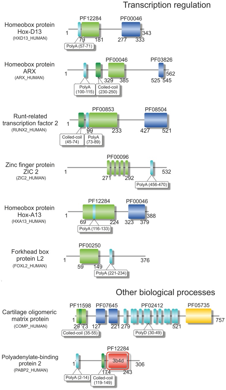
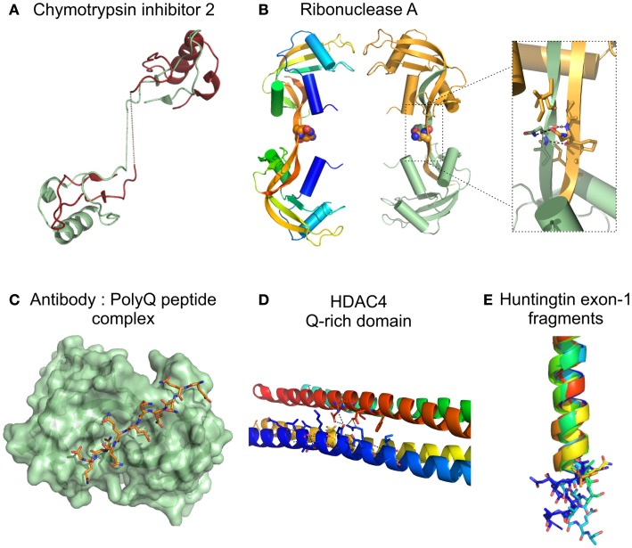
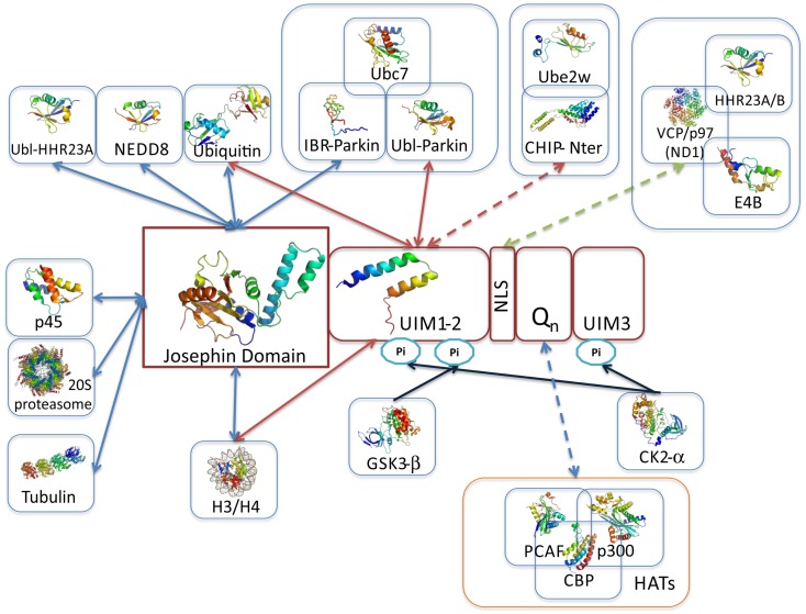
Trinucleotide repeat (TNR) expansions are present in a wide range of genes involved in several neurological disorders, being directly involved in the molecular mechanisms underlying pathogenesis through modulation of gene expression and/or the function of the RNA or protein it encodes. Structural and functional information on the role of TNR sequences in RNA and protein is crucial to understand the effect of TNR expansions in neurodegeneration. Therefore, this review intends to provide to the reader a structural and functional view of TNR and encoded homopeptide expansions, with a particular emphasis on polyQ expansions and its role at inducing the self-assembly, aggregation and functional alterations of the carrier protein, which culminates in neuronal toxicity and cell death. Detail will be given to the Machado-Joseph Disease-causative and polyQ-containing protein, ataxin-3, providing clues for the impact of polyQ expansion and its flanking regions in the modulation of ataxin-3 molecular interactions, function, and aggregation.

Keywords: amino acid-repeats; amyloid; microsatellites; protein aggregation; protein complexes; protein structure.


Figures

Figure 1
Figure 1
Structural variability of proteins encoded by TNR-containing genes. Illustrative domain graphics of the multi-domain structure of proteins associated with polyQ-expansion diseases. All proteins shown are referenced by their name as annotated in UniProt. The protein domains for which information is annotated in the Pfam database are shown as colored boxes with Pfam family accession code referenced above the domain box. Complete names of domains can be assessed by searching the specific Pfam accession code at http://pfam.sanger.ac.uk/. Numbers below the domain schemes represent amino acid residue numbers. Regions containing the amino acid repeats and with a prediction for formation of coiled-coils (as annotated in UniProt) are shown as well as regions with known 3D structure (boxed in red, with PDB accession codes shown). Notice the predominant location of the repeat regions within the N-terminal regions of the proteins.
Figure 2
Figure 2
Structural variability of proteins encoded by TNR-containing genes. Illustrative domain graphics of the multi-domain structure of proteins associated with polyD- and polyA-expansion diseases. All proteins shown are referenced by their name as annotated in UniProt. The protein domains for which information is annotated in the Pfam database are shown as colored boxes with Pfam family accession code referenced above the domain box. Complete names of domains can be assessed by searching the specific Pfam accession code at http://pfam.sanger.ac.uk/. Numbers below the domain schemes represent amino acid residue numbers. Regions containing the amino acid repeats and with a prediction for formation of coiled-coils (as annotated in UniProt) are shown as well as regions with known 3D structure (boxed in red, with PDB accession codes shown). Notice the predominant location of the repeat regions within the N-terminal regions of the proteins.
Figure 3
Figure 3
Structure of proteins/protein domains containing polyQ regions. (A) Cartoon representation of the domain swapped dimer of chymotrypsin inhibitor 2 with a 4 glutamine insertion [(Chen et al., 1999); PDB accession code 1cq4], dotted lines represent the polyQ linker not visible in the X-ray crystal structure. (B) Cartoon representation of domain swapped major dimers of ribonuclease A. Inset shows a short segment resembling the polar zipper formed by asparagine residues in the linker region [(Liu et al., 2001); PDB accession code 1f0v]. (C) Surface representation Fv fragment of a monoclonal antibody in complex with a polyQ peptide shown as sticks [(Li et al., 2007a), PDB accession code 2otu]. (D) Cartoon representation of the glutamine-rich domain from HDAC4 showing details of the polar interactions (dotted lines) at the oligomer interfaces involving glutamine residues [(Guo et al., 2007), PDB accession code 2o94]. (E) Cartoon representation of the crystal structures of huntingtin exon-1 fragments observed in different crystal forms, highlighting the different orientations of the C-terminal polyQ residues shown as sticks. The 17 glutamine stretch adopts variable conformations in the structures: α helix, random coil, and extended loop. [(Kim et al., 2009), PDB accession codes 3io4, 3iow, 3iov, 3iou, 3iot, 3ior, 3io6].
Figure 4
Figure 4
Overview of ataxin-3 structural information. Schematic illustration of ataxin-3 (isoform 2; a.k.a. 3UIM isoform) domain structure highlighting the regions involved in protein–protein interactions. The solution structures of the Josephin domain (PDB accession code 1yzb) and UIMs1-2 (PDB accession code 2klz) are shown colored from N-(blue) to C- terminus (red). JD-, UIM-, NLS-, and polyQ-mediated interactions are represented by blue, red, green, and purple arrows, respectively; blue arrows indicate the location of post-translational modification sites, resulting from the interaction and phosphorylation by CK2 and GSK3. Representative multi-subunit complexes where ataxin-3 participates are boxed (Li et al., ; Matsumoto et al., ; Scaglione et al., ; Durcan et al., 2012). One of the main questions in the quest for ataxin-3 interacting proteins is whether polyQ-expansion of the disease-protein modulates the binding affinities. Current data indicates that polyQ-expansion increments the ataxin-3 affinity for CHIP (Scaglione et al., 2011), VCP/p97 (Matsumoto et al., ; Boeddrich et al., ; Zhong and Pittman, 2006), and the transcription regulators p300, CBP, and PCAF (Li et al., 2002) (interactions represented by broken lines). Strikingly, all these interactions are mediated by ataxin-3 flexible tail, which includes the polyQ tract. Moreover the transcriptional regulators p300, CBP, and NCOR all contain amino acid repeats.
Figure 5
Figure 5
Overview of ataxin-3 protein interaction network. Data on the ataxin-3 interactors was obtained by analysis of Interactome3D (Mosca et al., 2012), MINT (Ceol et al., 2010), and Dr. PIAS (Sugaya and Furuya, 2011) protein interaction databases, and completed with data compiled from current literature on ataxin-3 protein associations obtained with a diverse set of experimental approaches (see complete information on Table 2). Red arrows indicate interactions for which structural data has been obtained, while orange arrows indicate that biophysical data on interaction affinity in vitro is known (Table 2). Broken arrows represent interactions that result from high-throughput interactome analysis that still require detailed biochemical and functional analysis. Proteins are grouped according to their biological role.

References

    1. Alba M. M., Guigo R. (2004). Comparative analysis of amino acid repeats in rodents and humans. Genome Res. 14, 549–55410.1101/gr.1925704 - DOI - PMC - PubMed
    1. Albrecht M., Golatta M., Wullner U., Lengauer T. (2004). Structural and functional analysis of ataxin-2 and ataxin-3. Eur. J. Biochem. 271, 3155–317010.1111/j.1432-1033.2004.04245.x - DOI - PubMed
    1. Altschuler E. L., Hud N. V., Mazrimas J. A., Rupp B. (1997). Random coil conformation for extended polyglutamine stretches in aqueous soluble monomeric peptides. J. Pept. Res. 50, 73–7510.1111/j.1399-3011.1997.tb00622.x - DOI - PubMed
    1. Amiel J., Trochet D., Clement-Ziza M., Munnich A., Lyonnet S. (2004). Polyalanine expansions in human. Hum. Mol. Genet. 2, R235–24310.1093/hmg/ddh251 - DOI - PubMed
    1. Antony P. M., Mantele S., Mollenkopf P., Boy J., Kehlenbach R. H., Riess O., et al. (2009). Identification and functional dissection of localization signals within ataxin-3. Neurobiol. Dis. 36, 280–29210.1016/j.nbd.2009.07.020 - DOI - PubMed

LinkOut - more resources