. 2013 Jun 20:4:158.
doi: 10.3389/fimmu.2013.00158. eCollection 2013.

Control of uterine microenvironment by foxp3(+) cells facilitates embryo implantation

Affiliations

Control of uterine microenvironment by foxp3(+) cells facilitates embryo implantation

Ana Teles et al. Front Immunol. .

Abstract

Implantation of the fertilized egg into the maternal uterus depends on the fine balance between inflammatory and anti-inflammatory processes. Whilst regulatory T cells (Tregs) are reportedly involved in protection of allogeneic fetuses against rejection by the maternal immune system, their role for pregnancy to establish, e.g., blastocyst implantation, is not clear. By using 2-photon imaging we show that Foxp3(+) cells accumulated in the mouse uterus during the receptive phase of the estrus cycle. Seminal fluid further fostered Treg expansion. Depletion of Tregs in two Foxp3.DTR-based models prior to pairing drastically impaired implantation and resulted in infiltration of activated T effector cells as well as in uterine inflammation and fibrosis in both allogeneic and syngeneic mating combinations. Genetic deletion of the homing receptor CCR7 interfered with accumulation of Tregs in the uterus and implantation indicating that homing of Tregs to the uterus was mediated by CCR7. Our results demonstrate that Tregs play a critical role in embryo implantation by preventing the development of a hostile uterine microenvironment.

Keywords: fibrosis; implantation; inflammation; pregnancy; regulatory T cells.


Figures
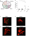
Figure 1
Figure 1
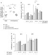
Tregs fluctuate during estrus cycle peaking at estrus, time point of sexual receptivity. Determination of the estrus cycle in non-pregnant females according to cell types in vaginal smears (A). Percentage of CD4+Foxp3GFP+ cells of female Foxp3GFP mice (n = 5/cycle stage) was analyzed in vaginal lavage (B). Data are expressed as single dot plots with medians and were analyzed by Mann–Whitney-U test (*P ≤ 0.05). Snap shots of two-photon microscopy videos of Foxp3GFP positive cells in uterus performed at different phases of the cycle (C) estrus phase; (D) diestrus phase; (E) metestrus phase; (F) proestrus phase.
Figure 2
Figure 2
Tregs expand in vivo and in vitro in presence of seminal fluid. Percentage of CD4+Foxp3+ cells in the uterine draining lymph nodes of CBA/J females mated with seminal vesicle-deficient, vasectomized, or intact BALB/c males was analyzed at day of conception (day 0.5) and compared to virgin CBA/J females (n = 4–7) (A). Statistical analysis were performed by Mann–Whitney-U test (**P ≤ 0.01). (B) Tregs were isolated from non-pregnant CBA/J females by magnetic cell sorting, stained with CFSE and cultured with seminal vesicle fluid (SVF) from BALB/c males. TGF-β1 was blocked with anti- TGF-β1 antibody, and proliferation of Treg determined after 24 h by using FACScan Calibur. Data are representative of four experiments and expressed as mean with SEM. Analysis was performed by two-way ANOVA test (**P ≤ 0.01). (C) Conventional T effector cells were isolated from non-pregnant CBA/J females by magnetic cell sorting, stained with CFSE and cultured with seminal vesicle fluid (SVF) from BALB/c males. TGF-β1 was blocked with anti- TGF-β1 antibody, and proliferation of Treg determined after 24 h by using FACScan Calibur. Data are representative of four experiments and expressed as mean with SEM. Analysis was performed by two-way ANOVA test and no statistically significant differences were found among the groups.
Figure 3
Figure 3
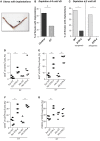
Embryo implantation is impaired after Treg depletion. Foxp3+ Treg were depleted in Foxp3DTR mice by application of DT every fourth day, starting 9 days before mating (day 9) with BALB/c males. Control groups received PBS. (A) shows a representative picture of a uterus stained at day 5 post conception with Chicago Blue dye application. The arrow indicates a representative implantation site. The percentage of females presenting implantations was analyzed on day 5 after mating [(B), n = 5–12]. Foxp3+ Treg were depleted in Foxp3.LuciDTR-4 mice by daily application of DT, starting on day 2 with allogeneic (CBAJ) or syngeneic (C57/BL6) males. Control groups were wt C57/BL6 females treated with DT. Implantation numbers were measured at day 5 [(C), n = 15–21]. For (B,C), data are expressed as medians of % of implanted females and analyzed by Fisher’s exact test (#P < 0.1, *P < 0.05). Not all plugged animals became pregnant. In samples from animals shown in Figure 3B, the percentage of CD8+ cells was analyzed in the uterus by flow cytometry (D), and the percentage of KI67+, CD44+, and CD62L+ (E–G) determined in uterine draining lymph nodes (n = 5/group). Data are expressed as medians and were analyzed by Mann–Whitney-U test (*P < 0.05; **P < 0.01).
Figure 4
Figure 4
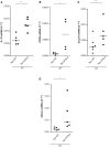
Depletion of Tregs creates an inflammatory uterine milieu. In samples from Foxp3.LuciDTR-4 or wild type animals treated with DT by daily application starting on day 2 and further mated syngeneically, inflammation markers were measured (n = 4–6/group) in uterine tissue by qPCR. Levels of IL-15 (A), CCR5 (B), CCL19 (C), and CXCL3 (D) were significantly elevated in mice depleted of Tregs as compared to DT-treated controls. Data are expressed as single dots with medians and were analyzed by Mann–Whitney-U test (#P < 0.1, P < 0.05; **P < 0.01).
Figure 5
Figure 5
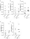
Depletion of Tregs does not provoke changes in the levels of IL-1b, gp130, TNF-α, ROR-γτ, or CCL5. In samples from Foxp3.LuciDTR-4 or wild type animals treated with DT by daily application starting on day 2 and further mated allogeneically, inflammation markers were measured (n = 4–6/group) in uterine tissue by qPCR. Levels of IL-1b (A), gp130 (B), TNF-α (C), RORγτ (D), and CCL5 (E) mRNA were comparable in mice depleted of Tregs and DT-treated controls. Data are expressed as single dot plots with medians and were analyzed by Mann–Whitney-U test. No statistically significant differences were observed between both groups in any of the molecules.
Figure 6
Figure 6
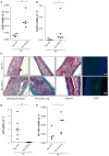
Treg depletion leads to uterine fibrosis. In samples from Foxp3.LuciDTR-4 or wild type animals treated with DT by daily application starting on day 2 and further mated syngeneically, fibrosis markers were measured (n = 4–6/group) in uterine tissue by qPCR. Levels of CtGF (A) and CXCL9 (B) were significantly elevated in mice depleted of Tregs as compared to DT-treated controls. Data are expressed as single dots with medians and were analyzed by Mann–Whitney-U test (#P < 0.1, **P < 0.01). Additionally, staining with Hematoxylin/Eosin revealed fibrosis areas with disorganized collagen fibers in animals without Tregs compared to wild type controls (C ii vs. i). Pricosirius red staining revealed a higher amount of green/blue-stained thin collagen fibers that confirms fibrosis in mice devoid of Tregs vs. controls (C iv vs. iii). Further, Masson’s staining manifested larger areas of collagen fibers in blue/violet in DT-treated Foxp3.Luci.DTR animals compared to DT-treated controls (C vi vs. v). Finally in (C vii, viii), immunofluorescence staining for CtGF confirmed accumulation of CtGF positive cells in mice depleted in Tregs as compared to the controls. All pictures were taken with a 10× objective. Analysis of uPA mRNA revealed that animals depleted in Tregs presented very low, almost undetectable levels of this molecule as compared to DT-treated controls (D). Treg depletion further provoked an augmentation of prostaglandin 1A mRNA (E).
Figure 7
Figure 7
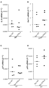
Treg depletion does not provoke changes in IL-9, Gal-1, LIF, or p53. In samples from Foxp3.LuciDTR-4 or wild type animals treated with DT by daily application starting on day 2 and further mated allogeneically, inflammation markers were measured (n = 4–6/group) in uterine tissue by qPCR. Levels of IL-9 (A), Gal-1 (B), LIF (C), and p53 (D) mRNA were comparable in mice depleted of Tregs and DT-treated controls. Data are expressed as single dot plots with medians and were analyzed by Mann–Whitney-U test. No statistically significant differences were observed between both groups in any of the molecules.

Similar articles

Cited by

References

    1. Ashkar A. A., Di Santo J. P., Croy B. A. (2000). Interferon gamma contributes to initiation of uterine vascular modification, decidual integrity, and uterine natural killer cell maturation during normal murine pregnancy. J. Exp. Med. 192, 259–27010.1084/jem.192.2.259 - DOI - PMC - PubMed
    1. Braundmeier A., Jackson K., Hastings J., Koehler J., Nowak R., Fazleabas A. (2012). Induction of endometriosis alters the peripheral and endometrial regulatory T cell population in the non-human primate. Hum. Reprod. 27, 1712–172210.1093/humrep/des083 - DOI - PMC - PubMed
    1. Carmeliet P., Schoonjas L., Kieckens L., Ream B., Degen J., Bronson R., et al. (1994). Physiological consequences of loss of plasminogen activator gene function in mice. Nature 368, 419–42410.1038/368419a0 - DOI - PubMed
    1. Fontenot J. D., Rasmunssen J. P., Gavin M. A., Rudensky A. Y. (2005). A function for interleukin-2 in Foxp3-expressing regulatory T cells. Nat. Immunol. 6, 1142–115110.1038/ni1263 - DOI - PubMed
    1. Gressner O. A., Gressner A. M. (2008). Connective tissue growth factor: a fibrogenic master switch in fibrotic liver diseases. Liver Int. 28, 1065–107910.1111/j.1478-3231.2008.01826.x - DOI - PubMed