

Quantitative Immunohistochemistry of Desmosomal Proteins (Plakoglobin, Desmoplakin and Plakophilin), Connexin-43, and N-cadherin in Arrhythmogenic Cardiomyopathy: An Autopsy Study
- PMID: 23802019
- PMCID: PMC3680985
- DOI: 10.2174/1874192401307010028
Quantitative Immunohistochemistry of Desmosomal Proteins (Plakoglobin, Desmoplakin and Plakophilin), Connexin-43, and N-cadherin in Arrhythmogenic Cardiomyopathy: An Autopsy Study
Abstract
Background: Arrhythmogenic right ventricular cardiomyopathy (ARVC) is a genetic disorder related to mutations in desmosomal proteins. The current study tests the hypothesis that immunohistochemical staining for desmosomal proteins is of diagnostic utility by studying autopsy-confirmed cases of ARVC.
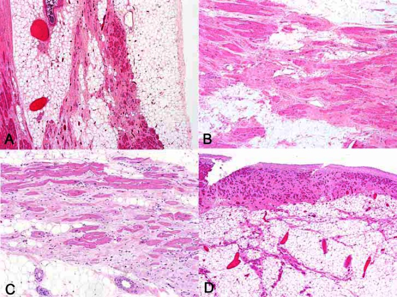
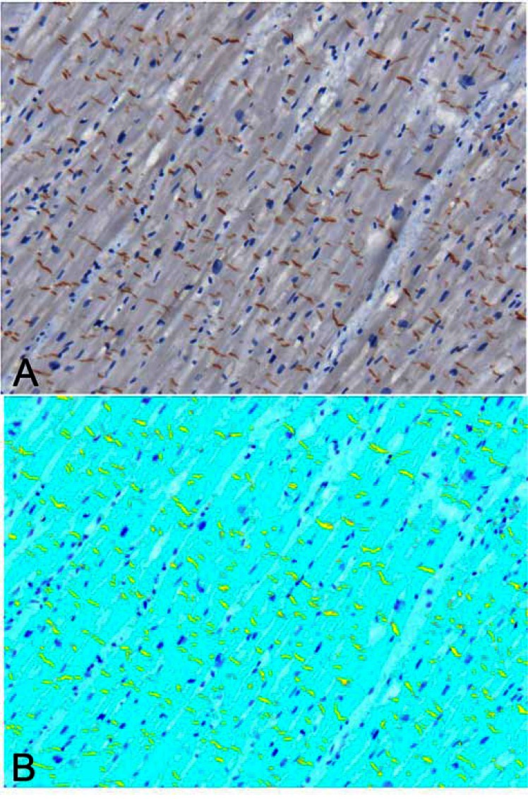
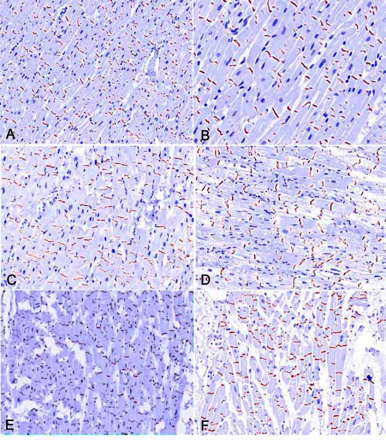
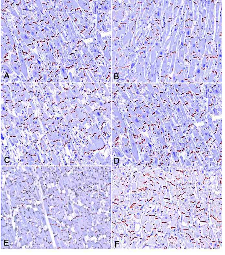
Methods and results: We studied 23 hearts from patients dying suddenly with ARVC. Control subject tissues were 21 hearts from people dying from non-cardiac causes (n=15), dilated cardiomyopathy (n=3) and coronary artery disease (n=3). Areas free of fibrofatty change or scarring were assessed on 50 sections from ARVC (24 left ventricle, 26 right ventricle) and 28 sections from controls. Immunohistochemical stains against plakoglobin, plakophilin, desmoplakin, connexin-43, and N-cadherin were applied and area expression analyzed by computerized morphometry. Desmin was stained as a control for fixation and similarly analyzed. The mean area of desmin expression was similar in controls and ARVC (86% vs. 85%, p=0.6). Plakoglobin expression was 4.9% ± 0.3% in controls, vs. 4.6% ± 0.3% in ARVC (p=0.3). Plakophilin staining was 4.8% ± 0.3% in controls vs. 4.4% ± 03% in ARVC (p=0.3). Desmoplakin staining was 3.4% in controls vs. 3.2 ± 0.2% in ARVC (p=0.6). There were no significant differences when staining was compared between right and left ventricles (all p > 0.1). For non-desmosomal proteins, the mean area of connexin-43 staining showed no significant difference by presence of disease.
Conclusions: The small and insignificant decrease in junction protein expression in ARVC suggests that immunohistochemistry is not a useful tool for the diagnosis.
Keywords: ARVC; arrhytmogenic cardiomyopaty; autopsy.; sudden death.
Figures
References
-
- Dalal D, Molin LH, Piccini J, et al. Clinical features of arrhythmogenic right ventricular dysplasia/cardiomyopathy associated with mutations in plakophilin-2. Circulation. 2006;113:1641–9. - PubMed
-
- Gerull B, Heuser A, Wichter T, et al. Mutations in the desmosomal protein plakophilin-2 are common in arrhythmogenic right ventricular cardiomyopathy. Nat Genet. 2004;36:1162–4. - PubMed
-
- Pilichou K, Nava A, Basso C, et al. Mutations in desmoglein-2 gene are associated with arrhythmogenic right ventricular cardiomyopathy. Circulation. 2006;113:1171–9. - PubMed
-
- Syrris P, Ward D, Asimaki A, et al. Desmoglein-2 mutations in arrhythmogenic right ventricular cardiomyopathy: a genotype-phenotype characterization of familial disease. Eur Heart J. 2007;28:581–8. - PubMed
LinkOut - more resources
Full Text Sources
Other Literature Sources
Research Materials