

Morphological and Functional Parameters in Patients with Tooth Wear before and after Treatment
- PMID: 23802024
- PMCID: PMC3681002
- DOI: 10.2174/1874210601307010055
Morphological and Functional Parameters in Patients with Tooth Wear before and after Treatment
Abstract
Advanced tooth wear often results in lost vertical dimension and impacts facial aesthetics. Complex restorative treatment can replace the lost tooth structure and improve functional occlusal and facial skeleton parameters.
Purpose: The aim of the study is to assess changes in the morphological and functional occlusal parameters of the facial skeleton after prosthetic rehabilitation that increased lost occlusal vertical dimension.
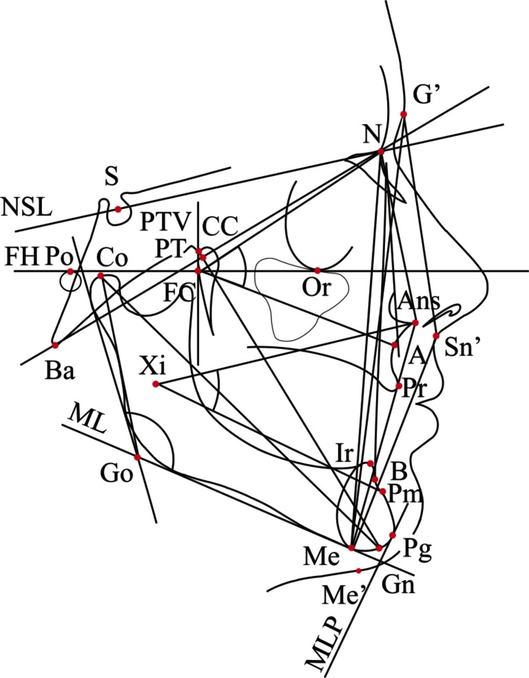
Material and methodology: 50 patients with advanced tooth wear were clinically examined, to assess the degree of wear. Each subject underwent cephalometric analysis, digital occlusal analysis, and electromyographic analysis, of the anterior temporalis, superficial masetter, anterior digastric, and the sternocleidomastoid muscles. Prosthodontic treatment was performed to restore the occlusal vertical dimension of each subject's occlusion, which was followed by repeating the pretreatment analyses. Pre and post treatment parameters were statistically compared.
Results: Pre-treatment cephalometric analysis showed that lost vertical dimension reduced anterior facial height and resulted in small angular skeletal parameters. Post treatment anterior facial height increased from the increased occlusal vertical dimension. The mean value of functional electrical activity during clenching post treatment, increased compared to pretreatment.
Conclusion: Increasing the vertical dimension of occlusion improved facial aesthetics by positively affecting facial skeletal angles. The restored occlusal surface morphology changed the pre treatment flat broad occlusal contacts into more point contacts. The increased vertical dimension of occlusion after treatment also increased muscle activity levels over the pretreatment levels after three months period of adaptation.
Keywords: Cephalometric analysis; EMG; T-Scan.; occlusal vertical dimension; rehabilitation; tooth wear.
Figures
Similar articles
-
Assessment of masticatory muscle activity and occlusion time in patients with advanced tooth wear.Arch Oral Biol. 2015 Sep;60(9):1346-55. doi: 10.1016/j.archoralbio.2015.06.006. Epub 2015 Jun 22. Arch Oral Biol. 2015. PMID: 26126289
-
The facial effects of tooth wear rehabilitation as measured by 3D stereophotogrammetry.J Dent. 2018 Jun;73:105-109. doi: 10.1016/j.jdent.2018.04.014. Epub 2018 Apr 22. J Dent. 2018. PMID: 29689294
-
The association between dental wear and reduced vertical dimension of the face: a morphologic study on human skulls.Arch Oral Biol. 2015 Jan;60(1):174-80. doi: 10.1016/j.archoralbio.2014.09.016. Epub 2014 Oct 13. Arch Oral Biol. 2015. PMID: 25455132
-
Occlusal Vertical Dimension: Best Evidence Consensus Statement.J Prosthodont. 2021 Apr;30(S1):12-19. doi: 10.1111/jopr.13315. J Prosthodont. 2021. PMID: 33783090 Review.
-
[Application of cephalometric analysis for determination of vertical dimension of occlusion--a literature review].Med Pregl. 2012 May-Jun;65(5-6):217-22. doi: 10.2298/mpns1206217s. Med Pregl. 2012. PMID: 22730706 Review. Serbian.
Cited by
-
The Prevalence and Overlaps of Temporomandibular Disorders in Patients with Myofascial Pain with Referral-A Pilot Study.Int J Environ Res Public Health. 2021 Sep 18;18(18):9842. doi: 10.3390/ijerph18189842. Int J Environ Res Public Health. 2021. PMID: 34574764 Free PMC article.
-
Comparative Evaluation of Occlusion before and after Soft Tissue Mobilization in Patients with Temporomandibular Disorder-Myofascial Pain with Referral.Int J Environ Res Public Health. 2021 Jun 18;18(12):6568. doi: 10.3390/ijerph18126568. Int J Environ Res Public Health. 2021. PMID: 34207403 Free PMC article.
References
-
- Johansson A, Johansson AK, Omar R, Carlsson GE. Rehabilitation of the worn dentition. J Oral Rehabil. 2008;35:548–66. - PubMed
-
- Tallgren A. Changes in adult face height due to ageing, wear and loss of teeth and prosthetic treatment. Acta Odontol Scand. 1957;15(Supp 24):73.
-
- Nakai N, Abekura H, Hamada T, Morimoto T. Comparison of the most comfortable mandibular position with the intercuspal position using cephalometric analysis. J Oral Rehabil. 1998;25:370–5. - PubMed
-
- Bakke M, Michler L, Moller E. Occlusal control of mandibular elevator muscle. Scand J Dent Res. 1992;100:284–91. - PubMed
-
- Hidaka O, Iwasaki M, Saito M, Morimoto T. Influence of clenching intensity on bite force balance, occlusal contact area, and average bite pressure. J Dent Res. 1999;72:1336–44. - PubMed
LinkOut - more resources
Full Text Sources
Other Literature Sources