

Arthroscopic trans-portal deep medial collateral ligament pie-crusting release
- PMID: 23802093
- PMCID: PMC3691777
- DOI: 10.1016/j.eats.2012.10.008
Arthroscopic trans-portal deep medial collateral ligament pie-crusting release
Abstract
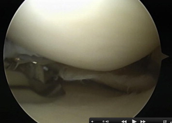
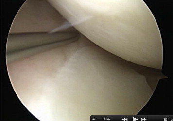
Arthroscopic treatments of meniscal injuries of the knee are among the most common orthopaedic procedures performed. Adequate visualization of the posterior horn of the medial meniscus might be challenging, especially in patients with tight medial compartments. In these cases instrument manipulation in an attempt to reach the posterior horn of the meniscus can cause an iatrogenic chondral injury because of the narrow medial joint space. A transcutaneous medial collateral ligament (MCL) pie-crusting release facilitates expansion of the medial joint space in a case of a tight medial compartment. Nevertheless, it might cause injury to the superficial MCL, infection, and pain and injury to the saphenous nerve because of multiple needle punctures of the skin. We describe an inside-out, arthroscopic deep MCL pie-crusting release, which allows access to the medial meniscus through the anterior approach to provide good visualization of the footprint and sufficient working space.
Figures


References
-
- Metcalf M.H., Barrett G.R. Prospective evaluation of 1485 meniscal tear patterns in patients with stable knees. Am J Sports Med. 2004;32:675–680. - PubMed
-
- Garrett W.E., Jr., Swiontkowski M.F., Weinstein J.N. American Board of Orthopaedic Surgery Practice of the Orthopaedic Surgeon: Part-II, certification examination case mix. J Bone Joint Surg Am. 2006;88:660–667. - PubMed
-
- Renstrom P., Johnson R.J. Anatomy and biomechanics of the menisci. Clin Sports Med. 1990;9:523–538. - PubMed
-
- Ahn J.H., Wang J.H., Yoo J.C., Noh H.K., Park J.H. A pull out suture for transection of the posterior horn of the medial meniscus: Using a posterior trans-septal portal. Knee Surg Sports Traumatol Arthrosc. 2007;15:1510–1513. - PubMed
-
- Kim Y.M., Rhee K.J., Lee J.K., Hwang D.S., Yang J.Y., Kim S.J. Arthroscopic pullout repair of a complete radial tear of the tibial attachment site of the medial meniscus posterior horn. Arthroscopy. 2006;22:795.e1–795.e4. - PubMed
LinkOut - more resources
Full Text Sources
Other Literature Sources
Medical
