Sulfopeptide probes of the CXCR4/CXCL12 interface reveal oligomer-specific contacts and chemokine allostery
- PMID: 23802178
- PMCID: PMC3783652
- DOI: 10.1021/cb400274z
Sulfopeptide probes of the CXCR4/CXCL12 interface reveal oligomer-specific contacts and chemokine allostery
Abstract
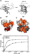
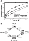
Tyrosine sulfation is a post-translational modification that enhances protein-protein interactions and may identify druggable sites in the extracellular space. The G protein-coupled receptor CXCR4 is a prototypical example with three potential sulfation sites at positions 7, 12, and 21. Each receptor sulfotyrosine participates in specific contacts with its chemokine ligand in the structure of a soluble, dimeric CXCL12:CXCR4(1-38) complex, but their relative importance for CXCR4 binding and activation by the monomeric chemokine remains undefined. NMR titrations with short sulfopeptides showed that the tyrosine motifs of CXCR4 varied widely in their contributions to CXCL12 binding affinity and site specificity. Whereas the Tyr21 sulfopeptide bound the same site as in previously solved structures, the Tyr7 and Tyr12 sulfopeptides interacted nonspecifically. Surprisingly, the unsulfated Tyr7 peptide occupied a hydrophobic site on the CXCL12 monomer that is inaccessible in the CXCL12 dimer. Functional analysis of CXCR4 mutants validated the relative importance of individual CXCR4 sulfotyrosine modifications (Tyr21 > Tyr12 > Tyr7) for CXCL12 binding and receptor activation. Biophysical measurements also revealed a cooperative relationship between sulfopeptide binding at the Tyr21 site and CXCL12 dimerization, the first example of allosteric behavior in a chemokine. Future ligands that occupy the sTyr21 recognition site may act as both competitive inhibitors of receptor binding and allosteric modulators of chemokine function. Together, our data suggests that sulfation does not ubiquitously enhance complex affinity and that distinct patterns of tyrosine sulfation could encode oligomer selectivity, implying another layer of regulation for chemokine signaling.
Figures


References
-
- Zou YR, Kottmann AH, Kuroda M, Taniuchi I, Littman DR. Function of the chemokine receptor CXCR4 in haematopoiesis and in cerebellar development. Nature. 1998;393:595–599. - PubMed
-
- Sierro F, Biben C, Martinez-Munoz L, Mellado M, Ransohoff RM, Li M, Woehl B, Leung H, Groom J, Batten M, Harvey RP, Martinez AC, Mackay CR, Mackay F. Disrupted cardiac development but normal hematopoiesis in mice deficient in the second CXCL12/SDF-1 receptor, CXCR7. Proc. Natl. Acad. Sci. U. S. A. 2007;104:14759–14764. - PMC - PubMed
-
- Burns JM, Summers BC, Wang Y, Melikian A, Berahovich R, Miao Z, Penfold ME, Sunshine MJ, Littman DR, Kuo CJ, Wei K, McMaster BE, Wright K, Howard MC, Schall TJ. A novel chemokine receptor for SDF-1 and I-TAC involved in cell survival, cell adhesion, and tumor development. J. Exp. Med. 2006;203:2201–2213. - PMC - PubMed
Publication types
MeSH terms
Substances
Grants and funding
- R01 GM081763/GM/NIGMS NIH HHS/United States
- R37 AI058072/AI/NIAID NIH HHS/United States
- R56AI063325/AI/NIAID NIH HHS/United States
- R56 AI063325/AI/NIAID NIH HHS/United States
- R01GM081763/GM/NIGMS NIH HHS/United States
- T32 GM007752/GM/NIGMS NIH HHS/United States
- F32GM103005/GM/NIGMS NIH HHS/United States
- R01AI058072/AI/NIAID NIH HHS/United States
- R01GM097381/GM/NIGMS NIH HHS/United States
- T32 GM008326/GM/NIGMS NIH HHS/United States
- R01 GM097381/GM/NIGMS NIH HHS/United States
- F32 GM103005/GM/NIGMS NIH HHS/United States
- T32GM007752/GM/NIGMS NIH HHS/United States
- R01 AI037113/AI/NIAID NIH HHS/United States
- R01 AI058072/AI/NIAID NIH HHS/United States
- U01 GM094612/GM/NIGMS NIH HHS/United States
- U01GM094612/GM/NIGMS NIH HHS/United States
LinkOut - more resources
Full Text Sources
Other Literature Sources

