. 2014 Mar 20;20(9):1410-22.
doi: 10.1089/ars.2012.5134. Epub 2013 Aug 12.

Functional consequences of radiation-induced oxidative stress in cultured neural stem cells and the brain exposed to charged particle irradiation

Affiliations

Functional consequences of radiation-induced oxidative stress in cultured neural stem cells and the brain exposed to charged particle irradiation

Bertrand P Tseng et al. Antioxid Redox Signal. .

Abstract

Aims: Redox homeostasis is critical in regulating the fate and function of multipotent cells in the central nervous system (CNS). Here, we investigated whether low dose charged particle irradiation could elicit oxidative stress in neural stem and precursor cells and whether radiation-induced changes in redox metabolism would coincide with cognitive impairment.

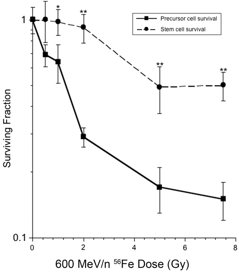
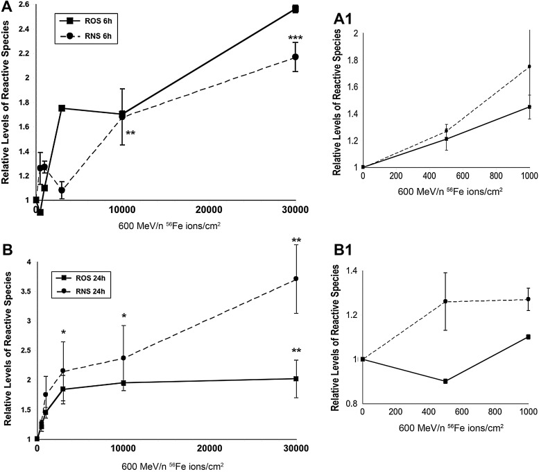
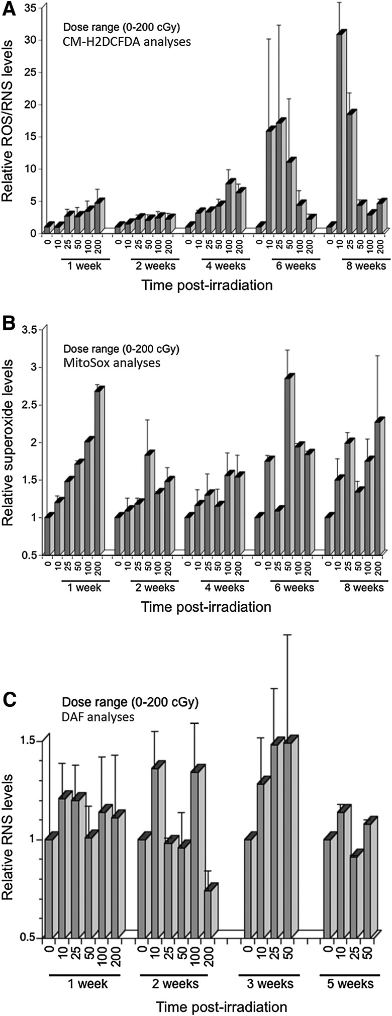
Results: Low doses (<1 Gy) of charged particles caused an acute and persistent oxidative stress. Early after (<1 week) irradiation, increased levels of reactive oxygen and nitrogen species were generally dose responsive, but were less dependent on dose weeks to months thereafter. Exposure to ion fluences resulting in less than one ion traversal per cell was sufficient to elicit radiation-induced oxidative stress. Whole body irradiation triggered a compensatory response in the rodent brain that led to a significant increase in antioxidant capacity 2 weeks following exposure, before returning to background levels at week 4. Low dose irradiation was also found to significantly impair novel object recognition in mice 2 and 12 weeks following irradiation.

Innovation: Data provide evidence that acute exposure of neural stem cells and the CNS to very low doses and fluences of charged particles can elicit a persisting oxidative stress lasting weeks to months that is associated with impaired cognition.

Conclusions: Exposure to low doses of charged particles causes a persistent oxidative stress and cognitive impairment over protracted times. Data suggest that astronauts subjected to space radiation may develop a heightened risk for mission critical performance decrements in space, along with a risk of developing long-term neurocognitive sequelae.


Figures

<b>FIG. 1.</b>
FIG. 1.
Cell survival and sphere formation following charged particle exposure. Neural stem and precursor cells subjected to iron ion irradiation were analyzed 5 days later for cell counts and sphere formation. The surviving fraction represents the number of cells (solid line) or spheres (dashed line) corrected for seeding density and normalized to unirradiated controls. Error bars represent the mean±S.E.M. of three to four independent measurements. *p<0.05; **p<0.001 for differences between precursor cells and spheres.
<b>FIG. 2.</b>
FIG. 2.
Dose-response for oxidative stress after low dose irradiation of neural stem and precursor cells with 600 MeV/n 56Fe ions. Neurospheres were irradiated from 1–15 cGy and analyzed for increased ROS/RNS using the redox-sensitive dye CM-H2DCFDA 12 and 24 h afterward. Compared to sham-irradiated controls, low dose exposure elicited a dose-responsive increase in oxidative stress at each postirradiation time. All data expressed as mean±S.E.M. of three to four independent observations and normalized to unirradiated controls set to unity. One-way ANOVA is significant (p<0.0001) and **p<0.001. ANOVA, analysis of variance; ROS, reactive oxygen species; RNS, reactive nitrogen species.
<b>FIG. 3.</b>
FIG. 3.
Low fluence response for the induction of ROS/RNS after charged particle irradiation. Neurospheres were subjected to 600 MeV/n Fe ions at low ion fluences (500–30,000 particles/cm2) and assayed for ROS/RNS using the redox-sensitive dyes CM-H2DCFDA and DAF, 6 h (A) and 24 h afterward (B). Lower fluence responses are shown at expanded scale (A1, B1). Low particle fluences were found to cause dose-responsive increases in oxidative stress at each of these early postirradiation times. All data expressed as mean±S.E.M. of three to four independent observations and normalized to unirradiated controls set to unity. One-way ANOVA is significant (p<0.0001, A and p<0.003, B) and *p<0.05; **p<0.01; ***p<0.001.
<b>FIG. 4.</b>
FIG. 4.
Low fluence response for the induction of ROS/RNS after charged particle irradiation. Neurospheres subjected to 600 MeV/n Fe ions at low fluences (500–30,000 particles/cm2) were assayed 36 and 48 h after irradiation for ROS/RNS using the redox-sensitive dyes CM-H2DCFDA (A) and DAF (B). At these longer postirradiation intervals, low particle fluences were still found to elicit increase in oxidative stress over unirradiated controls. At these longer times however, dose-responsive increases were less evident, and elevated nitric oxide was apparent at only the higher fluences levels (≤30,000 particles/cm2). All data expressed as mean±S.E.M. of three to four independent observations and normalized to unirradiated controls set to unity. ** p<0.05.
<b>FIG. 5.</b>
FIG. 5.
Persistent oxidative stress in neural precursors exposed to 600 MeV/n 56Fe ions. Cells exposed to low dose 56Fe ion irradiation were incubated with either CM-H2DCFDA (A), Mitosox (B), or DAF (C) and analyzed for persistent ROS/RNS, superoxide, or nitric oxide respectively at the indicated times. In each case a trend was observed for elevated oxidative species that persisted over the course of 5–8 weeks. All data were normalized to unirradiated controls set to unity. Error bars represent mean±S.E.M. of two to three measurements.
<b>FIG. 6.</b>
FIG. 6.
Antioxidant assessment in the irradiated brain. Following irradiation, brains were prepared for the measurement of antioxidant levels (A, B) and various enzymatic activities (C–H). Irradiation with 0.1 or 1.0 Gy led to a significant (p<0.0001) increase in GSH levels at 2 weeks, with a trend toward elevated GSSG at week 4 following irradiation (A, B). Catalase (C) and SOD (D–F) activities were increased significantly (to varying extents) 2 weeks after irradiation, but not at week 4. GPx and GST activities followed similar trends (G, H). GPx, glutathione peroxidase; GST, glutathione-S-transferase. p-values: *, 0.02; **, 0.07; ***, 0.04; ++, 0.0002; +++, 0.0001.
<b>FIG. 7.</b>
FIG. 7.
Novel object recognition of sham-irradiated and 56Fe-irradiated male mice. The DI shows reduced exploratory preference for the novel object over the familiar object 2-weeks after irradiation. N=8 mice/treatment. *p<0.05 (two-tailed t-test). DI, discrimination index.
<b>FIG. 8.</b>
FIG. 8.
Impaired recognition memory in mice detected 12 weeks after exposure to 16O or 48Ti particles. The NOR task was used to calculate a DI, which shows that all irradiated cohorts had reduced preference to explore the novel object compared to unirradiated controls. Controls (0 Gy) showed selective preference for the novel object (DI=46.64±5.3). Exposure to 600 MeV/n 16O particles reduced exploration of the novel object significantly (DI=15.7±4.1, p<0.05 at 5 cGy; DI=14.78±11.8, p<0.05 at 30 cGy). Similarly, exposure to 500 MeV/n 48Ti particles was also found to significantly reduce exploration for the novel object (DI=23.74±12.8, p<0.05 at 5 cGy; DI=16.4±6.6, p<0.05 at 30 cGy).

References

    1. Acharya MM, Lan ML, Kan VH, Patel NH, Giedzinski E, et al. Consequences of ionizing radiation-induced damage in human neural stem cells. Free Radic Biol Med 49: 1846–1855, 2010 - PubMed
    1. Azari H, Louis SA, Sharififar S, Vedam-Mai V, and Reynolds BA. Neural-colony forming cell assay: an assay to discriminate bona fide neural stem cells from neural progenitor cells. J Vis Exp pii: 2639, 2011 - PMC - PubMed
    1. Azzam EI, de Toledo SM, Raaphorst GP, and Mitchel RE. Low-dose ionizing radiation decreases the frequency of neoplastic transformation to a level below the spontaneous rate in C3H 10T1/2 cells. Radiat Res 146: 369–373, 1996 - PubMed
    1. Barker GR, Bird F, Alexander V, and Warburton EC. Recognition memory for objects, place, and temporal order: a disconnection analysis of the role of the medial prefrontal cortex and perirhinal cortex. J Neurosci 27: 2948–2957, 2007 - PMC - PubMed
    1. Barker GR. and Warburton EC. When is the hippocampus involved in recognition memory? J Neurosci 31: 10721–10731, 2011 - PMC - PubMed

Publication types

MeSH terms