. 2013 Aug;162(4):1867-80.
doi: 10.1104/pp.113.217604. Epub 2013 Jun 26.

A rice virescent-yellow leaf mutant reveals new insights into the role and assembly of plastid caseinolytic protease in higher plants

Affiliations

A rice virescent-yellow leaf mutant reveals new insights into the role and assembly of plastid caseinolytic protease in higher plants

Hui Dong et al. Plant Physiol. 2013 Aug.

Abstract

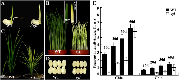
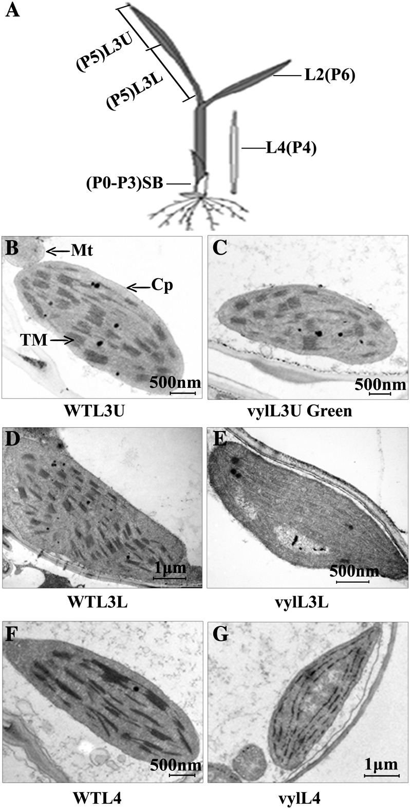
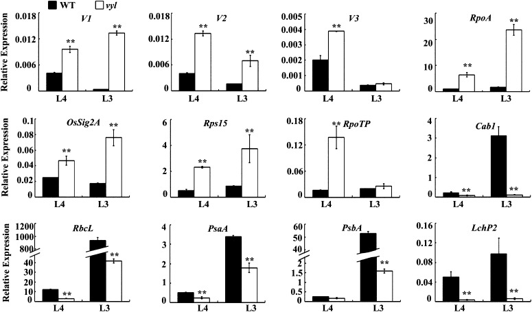
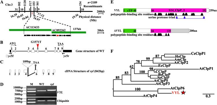
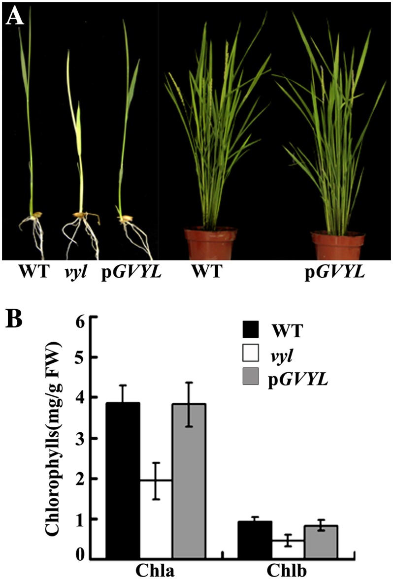
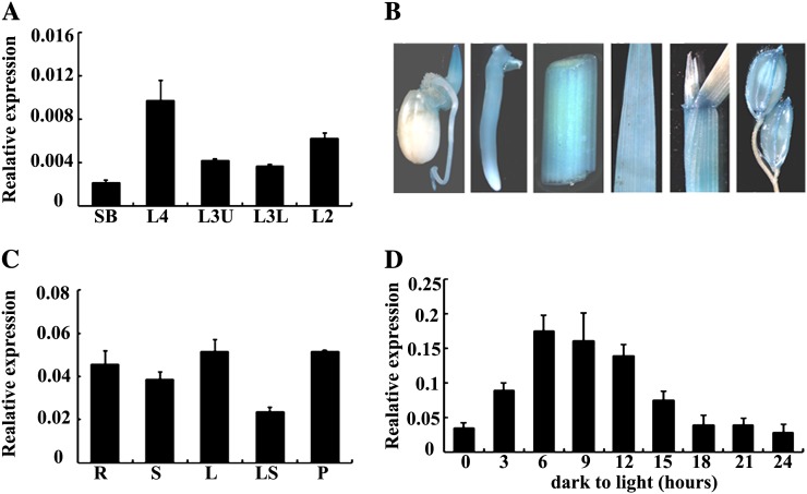
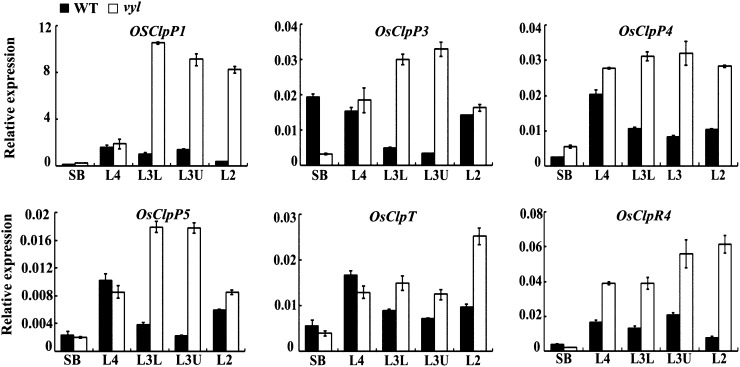
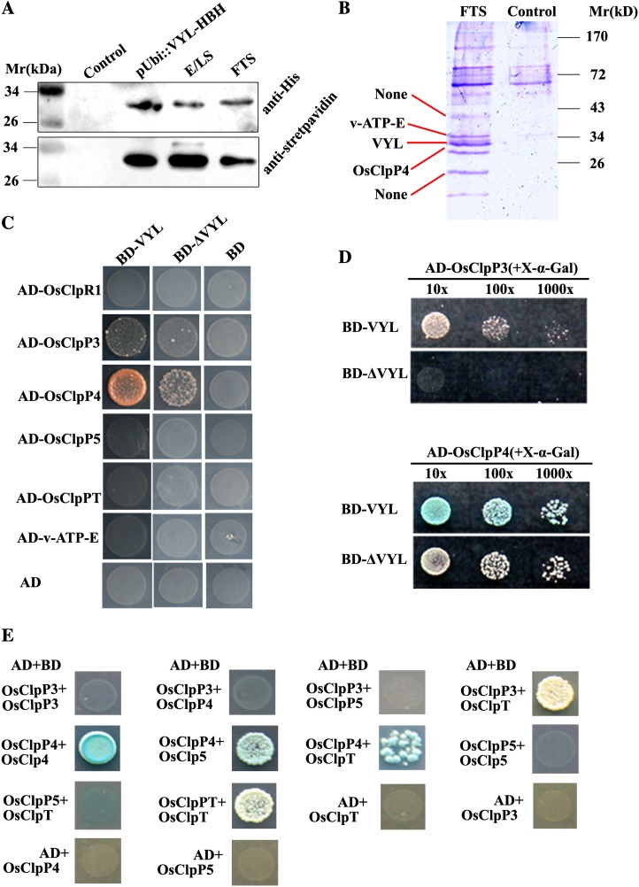
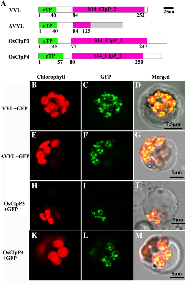
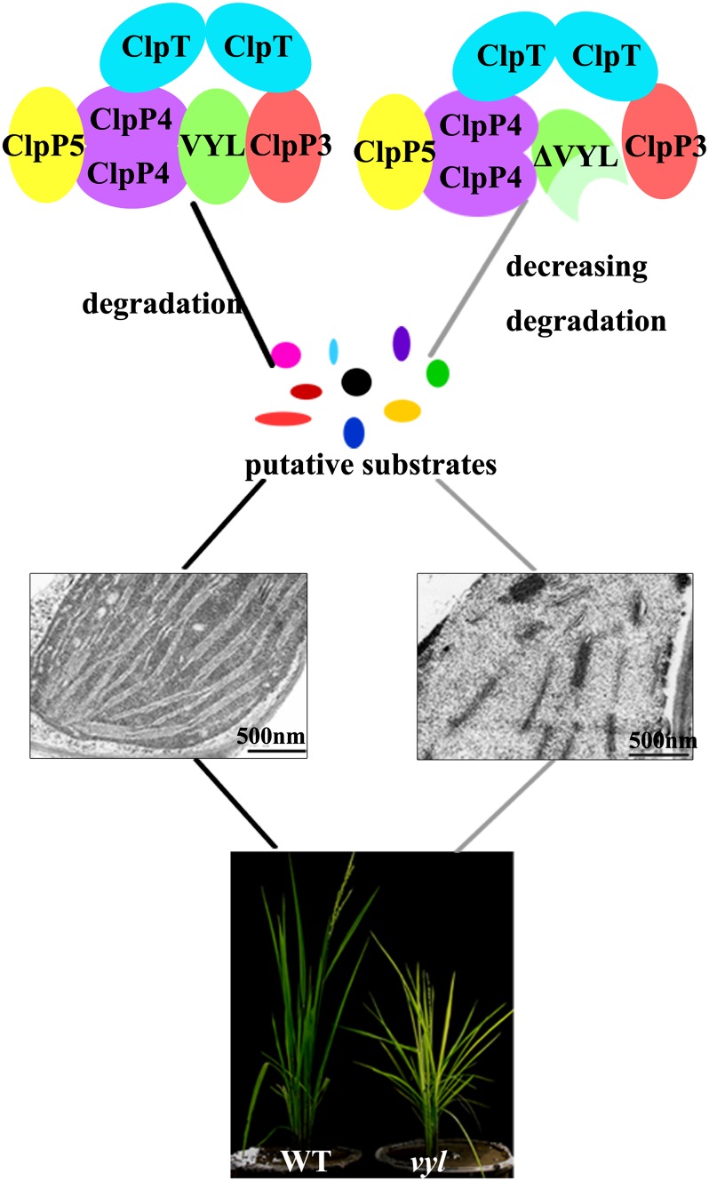
The plastidic caseinolytic protease (Clp) of higher plants is an evolutionarily conserved protein degradation apparatus composed of a proteolytic core complex (the P and R rings) and a set of accessory proteins (ClpT, ClpC, and ClpS). The role and molecular composition of Clps in higher plants has just begun to be unraveled, mostly from studies with the model dicotyledonous plant Arabidopsis (Arabidopsis thaliana). In this work, we isolated a virescent yellow leaf (vyl) mutant in rice (Oryza sativa), which produces chlorotic leaves throughout the entire growth period. The young chlorotic leaves turn green in later developmental stages, accompanied by alterations in chlorophyll accumulation, chloroplast ultrastructure, and the expression of chloroplast development- and photosynthesis-related genes. Positional cloning revealed that the VYL gene encodes a protein homologous to the Arabidopsis ClpP6 subunit and that it is targeted to the chloroplast. VYL expression is constitutive in most tissues examined but most abundant in leaf sections containing chloroplasts in early stages of development. The mutation in vyl causes premature termination of the predicted gene product and loss of the conserved catalytic triad (serine-histidine-aspartate) and the polypeptide-binding site of VYL. Using a tandem affinity purification approach and mass spectrometry analysis, we identified OsClpP4 as a VYL-associated protein in vivo. In addition, yeast two-hybrid assays demonstrated that VYL directly interacts with OsClpP3 and OsClpP4. Furthermore, we found that OsClpP3 directly interacts with OsClpT, that OsClpP4 directly interacts with OsClpP5 and OsClpT, and that both OsClpP4 and OsClpT can homodimerize. Together, our data provide new insights into the function, assembly, and regulation of Clps in higher plants.


Figures

Figure 1.
Figure 1.
Phenotypic comparison of wild-type (WT) and vyl plants. A, Phenotypes of 3-d-old wild-type and vyl seedlings. B, Phenotypes of 2-week-old wild-type and vyl seedlings. The inset shows the greening of the tip region of the second real leaf of the vyl plants. C, Phenotypes of wild-type and vyl plants at the heading stage showing reduced growth of the vyl mutant plants. D, Comparison of wild-type and vyl seeds (dehulled). E, Measurement of the chlorophyll content of all leaves of wild-type and vyl mutants at four different developmental stages: 10, 20, 30, and 60 d. Data are means ± sd (n = 5). Error bars represent sd of five independent experiments. Chla, Chlorophyll a; Chlb, chlorophyll b; fr. wt, fresh weight.
Figure 2.
Figure 2.
Ultrastructure of chloroplasts in mesophyll cells of 2-week-old wild-type (WT) and vyl plants. A, Diagram of a rice seedling with fully emerged third leaf. SB, Shoot base (approximately 8-mm piece from the bottom of the third leaf sheath). P0 to P6 indicate the six developmental stages of the leaf. Shoot base and L4 sections contain immature chloroplasts, while L3U, L3L, and L2 sections contain mature chloroplasts. B, D, and F, Electron micrographs of L3U, L3L, and L4, respectively, from wild-type plants with fully emerged third leaf. C, E, and G, Electron micrographs of L3U Green (the green section in L3U), L3L, and L4, respectively, from a vyl mutant with fully emerged third leaf. Chloroplasts of the wild type have well-ordered thylakoid and stacked membranes in L3U, L3L, and L4, but only chloroplasts of L3U Green (already turned green) from vyl mutants have normal stacked membranes. Chloroplasts of L4 and L3L from vyl mutants have much reduced thylakoid and stacked membranes. Cp, Chloroplast; Mt, mitochondria; TM, thylakoid membrane. Bars = 2 μm in B, C, E, and F and 500 nm in D and G.
Figure 3.
Figure 3.
qRT-PCR analysis of genes associated with chloroplast biogenesis in the wild type (WT) and the vyl mutant. The relative expression level of each gene in the L4 and L3U sections of wild-type and vyl mutant seedlings (where chloroplasts are in the second and third developmental stages, respectively) were analyzed by qRT-PCR and normalized using the Ubiquitin gene as an internal control. Data are means ± sd (n = 3). Asterisks indicate statistically significant differences compared with the wild type at P < 0.05 by Student’s t test.
Figure 4.
Figure 4.
Map-based cloning of VYL and phylogenetic analysis of VYL. A, The VYL locus was mapped to the short arm of chromosome 3 between the Indel markers F17 and I53 and was narrowed to a 134-kb genomic DNA region located on the bacterial artificial chromosome clones AC113433 and AC092262. The black arrows denote the 17 putative ORFs in the 134-kb genomic region. LOC_OsO3g29810 is shown as the red arrow. B, Genomic structure of LOC_Os03g29810. The mutation site is indicated by the red arrowhead (G1571T). WT, Wild type. C, Diagram of the mutant cDNA structure of LOC_Os03g29810. D, Semiquantitative RT-PCR analysis showing the sizes of the wild-type VYL and mutant vyl full-length cDNAs. E, Diagram of the wild-type VYL and mutant ΔVYL proteins. aa, Amino acids; cTP, chloroplast-targeting peptide. F, Phylogenetic analysis of VYL and its related proteins. VYL is most closely related to AtClpP6. Am, Apis mellifera; At, Arabidopsis; Bs, Bacillus subtilis; Ec, E. coli; C, Cyanophyta sp. PCC 7425; Ce, Caenorhabditis elegans; Hs, Homo sapiens; Se, Synechococcus elongatus. The red star denotes VYL.
Figure 5.
Figure 5.
The VYL complementation test. A, Complementation of the vyl mutant. Phenotypes of wild-type (WT), vyl mutant, and transgenic plants harboring the pGVYL transgene. The left panel shows 10-d-old seedlings, and the right panel shows plants at the heading stage. B, Pigment contents of leaves of 10-d-old wild-type, vyl mutant, and transgenic plants harboring the pGVYL plasmid. Data are means ± sd (n = 5). Error bars represent sd of five independent experiments. Chla, Chlorophyll a; Chlb, chlorophyll b; FW, fresh weight.
Figure 6.
Figure 6.
Expression analysis of VYL. A, qRT-PCR analysis showing that VYL expression peaks in the L4 section of wild-type plants with fully emerged third leaves. The VYL gene was normalized using a Ubiquitin gene as an internal control. Error bars represent sd of three independent experiments. Asterisk indicates statistical significance at P < 0.05 by Student’s t test. B, Histochemical staining shows that the pVYL::GUS reporter gene is ubiquitously expressed in young buds, young roots, stems, leaves, leaf shoots, and panicles. C, qRT-PCR analysis showing that VYL is expressed in different tissues of the wild-type plants at the heading stage and was normalized using a Ubiquitin gene as an internal control (n = 3). D, qRT-PCR analysis of the VYL gene during greening of etiolated seedlings. After growing in darkness for 10 d, etiolated wild-type rice seedlings were illuminated for 3, 6, 9, 12, 15, 18, 21, or 24 h. The relative VYL RNA levels increased along with the increased illumination time and peaked at 6 h after illumination started. After that, VYL expression decreased. Seedlings grown under continuous light or darkness were used as controls. The VYL gene was normalized using a Ubiquitin gene as an internal control. Data are means ± sd (n = 3).
Figure 7.
Figure 7.
Expression analysis of several Clp genes. qRT-PCR analysis shows the relative expression levels of OsClpP1, OsClpP3, OsClpP4, OsClpP5, OsClpT, and OsClpR4. Each gene was normalized using a Ubiquitin gene as an internal control. Data are means ± sd (n = 3). WT, Wild type.
Figure 8.
Figure 8.
Isolation of proteins that interact with VYL, and interactions between VYL, OsClpR1, OsClpP3, OsClpP4, OsClpP5, and OsClpT. A, Immunoblot analysis of VYL-HBH fusion proteins from total protein extracts of pUbi::VYL-HBH T1 transgenic plants, the eluate from Ni2+-Sepharose (E/LS; corresponding to the sample loaded on streptavidin-agarose), and the flow-through streptavidin-agarose (FTS). The wild-type nontransgenic plant extract was used as the control. B, Coomassie brilliant blue-stained SDS-PAGE gel showing the extra bands from the pUbi::VYL-HBH transgenic plants compared with the control sample (nontransgenic wild-type plants). The three protein bands indicated by the red lines were excised and subjected to MALDI-TOF analysis. NONE means that the protein bands did not get a good enough score to be confident (P < 0.05). C, Yeast two-hybrid assays showing that VYL interacts with OsClpP3 and OsClpP4. D, Yeast two-hybrid assays showing that the ΔVYL mutant protein has reduced interactions with OsClpP3 and OsClpP4 compared with the wild-type VYL protein. X-α-Gal was directly added to the solid yeast medium, and the same amount of yeast was used in each assay. The strength of the interaction was judged from the intensity of blue color and yeast growth density. E, Yeast two-hybrid assays showing that OsClpP3 interacts with OsClpT and OsClpP4 interacts with OsClpP5 and OsClpT. In addition, both OsClpP4 and OsClpT can interact with themselves.
Figure 9.
Figure 9.
Subcellular localization of VYL proteins in rice protoplasts. A, Diagrams of the wild-type VYL, ΔVYL mutant, OsClpP3, and OsClpP4 proteins. aa, Amino acids; cTP, chloroplast-targeting peptide. B to D, GFP signals of VYL-GFP fusion proteins localized in the chloroplasts of rice protoplasts. E to G, GFP signals of ΔVYL-GFP fusion proteins localized in the chloroplasts of rice protoplasts. H to J, GFP signals of OsClpP3-GFP fusion proteins localized in the chloroplasts of rice protoplasts. K to M, GFP signals of OsClpP4-GFP fusion proteins localized in the chloroplasts of rice protoplasts. Fluorescence signals were visualized using confocal laser scanning microscopy. Green fluorescence shows GFP, red fluorescence indicates chloroplast autofluorescence, and yellow fluorescence indicates images with the two types of fluorescence merged. Bars = 7.5 μm in B to D and 5 μm in E to M.
Figure 10.
Figure 10.
Model showing a role of the Clp in regulating chloroplast biogenesis and leaf development in rice. WT, Wild type.

Similar articles

Cited by

References

    1. Adam Z, Rudella A, van Wijk KJ. (2006) Recent advances in the study of Clp, FtsH and other proteases located in chloroplasts. Curr Opin Plant Biol 9: 234–240 - PubMed
    1. Archer EK, Bonnett HT. (1987) Characterization of a virescent chloroplast mutant of tobacco. Plant Physiol 83: 920–925 - PMC - PubMed
    1. Baena-González E, Aro EM. (2002) Biogenesis, assembly and turnover of photosystem II units. Philos Trans R Soc Lond B Biol Sci 357: 1451–1459, discussion 1459–1460 - PMC - PubMed
    1. Bruch EM, Rosano GL, Ceccarelli EA. (2012) Chloroplastic Hsp100 chaperones ClpC2 and ClpD interact in vitro with a transit peptide only when it is located at the N-terminus of a protein. BMC Plant Biol 12: 57–64 - PMC - PubMed
    1. Bukau B, Weissman J, Horwich A. (2006) Molecular chaperones and protein quality control. Cell 125: 443–451 - PubMed

Publication types

MeSH terms

LinkOut - more resources