. 2013:4:2086.
doi: 10.1038/ncomms3086.

Homeostatic regulation of dendritic dynamics in a motor map in vivo

Affiliations

Homeostatic regulation of dendritic dynamics in a motor map in vivo

Sandeep Kishore et al. Nat Commun. 2013.

Abstract

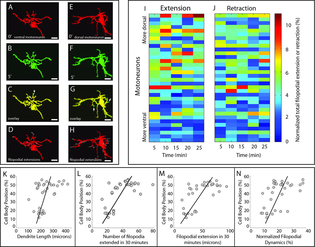
Neurons and circuits are remarkably dynamic. Their gross structure can change within minutes as neurons sprout and retract processes to form new synapses. Homeostatic processes acting to regulate neuronal activity contribute to these dynamics and predict that the dendritic dynamics within pools of neurons should vary systematically in accord with the activity levels of individual neurons in the pool during behaviour. Here we test this by taking advantage of a topographic map of recruitment of spinal motoneurons in zebrafish. In vivo imaging reveals that the dendritic filopodial dynamics of motoneurons map onto their recruitment pattern, with the most electrically active cells having the lowest dynamics. Genetic reduction of activity inverts this map of dynamics. We conclude that homeostatic mechanisms driven by a gradient of activity levels in a pool of neurons can drive an associated gradation in neuronal dendritic dynamics, potentially shaping connectivity within a functionally heterogenous pool of neurons.


Figures

Figure 1
Figure 1. Topographic organization of dendritic growth patterns
A, Schematic summarizing the age related dorsoventral organization of motoneurons in the spinal cord of zebrafish larvae. The youngest motoneurons are located in ventral spinal cord with older motoneurons occupying progressively more dorsal locations. This organization correlates with a gradient in the recruitment of motoneurons such that the youngest motoneurons are recruited most often during movements of all speeds, whereas the older, more powerful dorsal motoneurons are recruited less frequently at only high speeds. B, Images of motoneuron dendrites labeled with membrane tagged GFP (mGFP) at 2 days post fertilization (dpf) and 4 dpf. Right panel shows a motoneuron co-expressing PSD-95 (post synaptic density 95) tagged with GFP to mark glutamatergic synapses and a membrane targeted Cherry (mMCherry) to highlight the dendritic arbor. Arrows point to filopodia-like structures without PSD-95 puncta. C, Growth of motoneurons between 4 and 6 days. Left panel shows an overlay of the dendritic arbor of an individual motoneuron at 4 days (green) and 6 days (red). Middle panel shows the relationship between the dendritic length added between 4 and 6 days and the dorso-ventral position of the motoneuron. Right panel shows the dendritic growth between 4 and 6 days as a percentage of the total dendritic length at 4 days. Scale bars in B and C are 5 microns.
Figure 2
Figure 2. Dendritic dynamics vary systematically with soma position in spinal cord
A, B, Images of a ventral motoneuron expressing mGFP at the first two consecutive time points (0 min and 5 min). C, Overlaid image of A and B. Arrows point to filopodial extensions. D, Reconstruction (in white) of dendritic extensions over a 30 minute period in the same neuron. E,F, Images of a dorsal motoneuron at the first two consecutive time points (0 min and 5 min). G, Overlaid image of E and F. Arrows point to filopodial extensions. H, Reconstruction (in white) of the neuron’s filopodial extensions over a 30 minute period. I,J, Quantification of filopodial extensions and retractions between consecutive time points for 34 mGFP expressing motoneurons. Each row represents a single cell and the rows are arranged from bottom to top based on the ventral to dorsal position of the cell in the spinal cord. Each column represents the total length of filopodial extension between two consecutive time points normalized to the dendritic arbor length of that cell. Color map is on the right. K, Plot of cell body position versus length of dendritic arbor that remained unchanged over the entire imaging period for each motoneuron. Locations are normalized with respect to the dorsal (one) and ventral (zero) edges of spinal cord in all figures. L, Plot of cell body position versus total number of filopodia extended in the 30 minute imaging period. M, Plot of position versus the summed length of all filopodial extensions over the entire imaging period. N, Plot of soma position versus the normalized filopodial dynamics, which were calculated by dividing the sum of all filopodial extensions for a cell by the stable dendritic arbor length of that cell. Scale bars are 10 microns in A–H.
Figure 3
Figure 3. Kir2.1 expression increases dendritic dynamics in ventral motoneurons
A,B, Images of a ventral motoneuron expressing Kir2.1-viral2a-mGFP at the first two consecutive time points (0 min and 5 min). C, Overlaid image of A and B. Arrows point to filopodial extensions. D, Reconstruction (in white) of dendritic extensions over a 30 minute period. E,F, Images of a dorsal motoneuron at the first two consecutive time points (0 min and 5 min). G, Overlaid image of E and F. Arrows point to filopodial extensions. H, Reconstruction (in white) of filopodial extensions over a 30 minute period. I,J, Quantification of filopodial extensions and retractions between consecutive time points for 16 Kir2.1-viral2a-mGFP expressing motoneurons. Each row represents a single cell and the rows are stacked from bottom to top based on the ventral to dorsal position of the cell in the spinal cord. Each column represents the total length of filopodial extensions between two consecutive time points normalized to the dendritic arbor length of that cell. Color map is on the right. Scale bars are 10 microns in A–H.
Figure 4
Figure 4. Changing the excitability of motoneurons reverses the gradient of dendritic dynamics but does not affect stable dendritic size
A, Plot of cell body position versus total number of filopodia extended in the 30 minute imaging period for: 16 motoneurons expressing Kir2.1-viral2a-mGFP (red circles); 34 mGFP expressing motoneurons (gray circles); and 7 mutantKir2.1-viral2a-mGFP expressing motoneurons (blue circles). B, Plot of position versus the sum of all filopodial extensions over the entire imaging period. C, Plot of position versus filopodial dynamics normalized to the stable dendritic arbor length of the cell. D, Plot of cell body position versus stable dendritic arbor length for each group. E–G, Plots of retractions similar to those for extensions in A–D. H, Schematic showing the inverted gradient of motoneuron dendritic dynamics as a consequence of decreasing the excitability of motoneurons.

References

    1. Wu GY, Cline HT. Time-lapse in vivo imaging of the morphological development of Xenopus optic tectal interneurons. The Journal of comparative neurology. 2003;459:392–406. - PubMed
    1. Wong WT, Wong RO. Rapid dendritic movements during synapse formation and rearrangement. Current opinion in neurobiology. 2000;10:118–124. - PubMed
    1. Wong RO, Ghosh A. Activity-dependent regulation of dendritic growth and patterning. Nat Rev Neurosci. 2002;3:803–812. - PubMed
    1. Smith SJ. Dissecting dendrite dynamics. Science. 1999;283:1860–1861. - PubMed
    1. Holtmaat A, Svoboda K. Experience-dependent structural synaptic plasticity in the mammalian brain. Nat Rev Neurosci. 2009;10:647–658. - PubMed

Publication types

Substances

LinkOut - more resources