. 2013 Aug 16;288(33):23622-32.
doi: 10.1074/jbc.X113.497321. Epub 2013 Jun 26.

Chaperonin-mediated protein folding

Affiliations

Chaperonin-mediated protein folding

Arthur L Horwich. J Biol Chem. .

Abstract

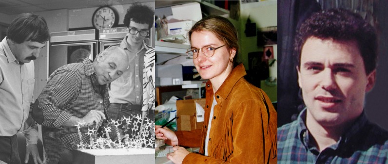
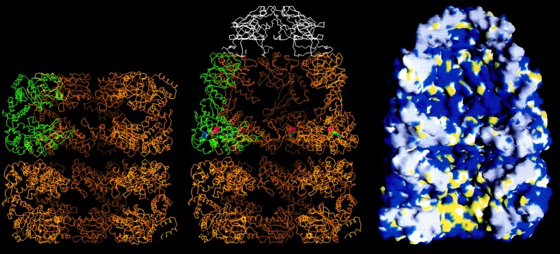
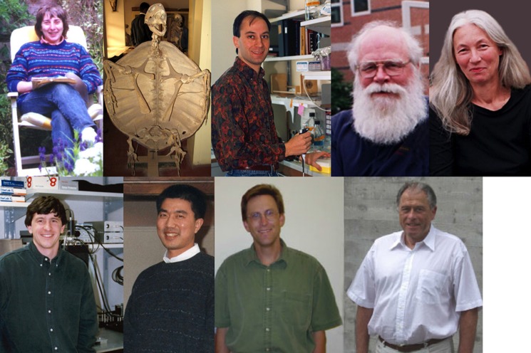
We have been studying chaperonins these past twenty years through an initial discovery of an action in protein folding, analysis of structure, and elucidation of mechanism. Some of the highlights of these studies were presented recently upon sharing the honor of the 2013 Herbert Tabor Award with my early collaborator, Ulrich Hartl, at the annual meeting of the American Society for Biochemistry and Molecular Biology in Boston. Here, some of the major findings are recounted, particularly recognizing my collaborators, describing how I met them and how our great times together propelled our thinking and experiments.

Keywords: Chaperone Chaperonin; Molecular Chaperone; Polypeptide; Protein Folding; Protein Misfolding; Yeast.


Figures

FIGURE 1.
FIGURE 1.
Imported mitochondrial proteins whose folding to the native form in the matrix compartment was deficient in our Hsp60-deficient yeast mutant. Ketoglutarate dehydrogenase (KGDH) and lipoamide dehydrogenase (LPDH) were observed to be affected in Ref. . Pre-existent functional Hsp60 was observed to be required for proper folding and subsequent assembly of newly imported Hsp60 in Ref. : no new Hsp60 complex could be produced in the mutant yeast after temperature shift. OM, outer membrane; IM, inner membrane; Fo SUIX, subunit 9 of F0-ATPase; F1β, F1-ATPase β-subunit; CS, citrate synthase. This figure was modified from Ref. .
FIGURE 2.
FIGURE 2.
Left, Ulrich Hartl in 1989, during a visit to his parents' home in the Black Forest. Food, wine, and ideas flowed freely. Middle, Ming Cheng in 1989. Right, Ming's impenetrable bench in the lab in 1989 (upper) and Ming's similarly occupied kitchen counter in Taipei in 2012 (lower).
FIGURE 3.
FIGURE 3.
Left, Andrzej Joachimiak (left), Paul Sigler (middle), and Zbyszek Otwinowski (right) in 1989, inspecting a crystallographic model of the trp repressor-operator complex, a structure they had recently solved. Middle, Kerstin Braig generated the first well diffracting crystals of GroEL in 1993. Right, David Boisvert, here in 1995, provided major impetus to the crystallization work. He worked on a monoclinic crystal form of GroEL and solved the structure of GroEL-ATPγS (work that was carried out with help from Jimin Wang). Note that many members of the Sigler lab, including Dan Gewirth, Greg Van Duyne, and Rashmi Hegde, also contributed to the GroEL structure determination, helping particularly with data collection and refinement. Refinement efforts ultimately relied heavily on the group of Axel Brunger (e.g. Ref. 41).
FIGURE 4.
FIGURE 4.
Cutaway images of models of GroEL alone (left) and complexed with nucleotide and GroES (middle and right). The left and middle images are Cα traces of the machine, with one of the seven subunits of GroEL colored green and the subunits of GroES colored white. In the image of GroEL alone (left), the equatorial domains are visible, forming the waistline of the cylinder, making slender covalent connections via the intermediate domains to the terminal apical domains, at either end of the cylinder. Note that the central cavity is blocked at the equatorial level of both rings by the collective of disordered C-terminal tails. The hydrophobic polypeptide-binding surface at the inside aspect of the apical domains of an open ring is colored yellow in the space filling model (right, bottom ring). When nucleotide and GroES bind, the intermediate and apical domains undergo rigid body movements that involve a downward rotation of the intermediate domain that locks the nucleotide into the bound ring (middle, red and blue sphere) and involves overall elevation and clockwise twisting movements of the apical domains that remove their hydrophobic surfaces from facing the cavity, ejecting the initially bound polypeptide into it, whereupon folding commences. Note that the ejected polypeptide resides in a hydrophilic chamber, denoted by blue in the cavity of the top ring (right). This figure is modified from Ref. .
FIGURE 5.
FIGURE 5.
Upper row, left to right, Helen Saibil, our long-time EM collaborator, pictured at home in London in her backyard reading area on a surprisingly sunny day; image of a tortoise skeleton (not a lab member) from one of Helen's travels to Ghana; Jonathan Weissman, circa 1995; Wayne Fenton, timeless; and Krystyna Furtak, who has been with me from day one and produced approximately one-hundred GroEL mutant constructs. Lower row, left to right: Hays Rye, who fluorescently labeled the reaction components and watched them come and go in real time; Zhaohui Xu, who crystallized GroEL-GroES-ADP; Eric Bertelsen, unafraid to design and produce the molecules enabling NMR approaches to the machinery; and Kurt Wüthrich, our long-time NMR collaborator and originator of TROSY techniques for studying large proteins.
FIGURE 6.
FIGURE 6.
GroEL-GroES reaction cycle. An open ring becomes rapidly occupied with ATP in the seven equatorial sites, with binding within a ring occurring cooperatively and between rings with negative cooperativity (42). This dictates an inherent asymmetry to the machine, as GroES can bind only to ATP-mobilized apical domains. Following rapid ATP binding, the polypeptide arrives, coming on at about one-tenth the rate of ATP over several hundred milliseconds (first panel) (43). Subsequently, GroES collides with the ATP-mobilized apical domains, forming a ternary collision complex in which both polypeptide and GroES are simultaneously bound to the apical domains, preventing any chance of polypeptide escape. Next, large and forceful rigid body elevation and twisting of the apical domains lead to release of the polypeptide into the encapsulated chamber, where the chain commences folding (second panel). This is the longest step of the reaction cycle, ∼10 s, following which ATP hydrolysis (third panel) weakens the affinity for GroES, and subsequent binding of ATP in the opposite ring sends an allosteric signal that ejects GroES, polypeptide, and hydrolyzed ADP from what had been the folding-active ring (fourth panel) (44). At the same time, the opposite ring is now set up to become the folding-active ring (fifth panel). T, ATP; D, ADP; N, natively folded protein; Ic, intermediate committed to the native state; Iuc, intermediate not committed to the native state. This figure is from Ref. .

References

    1. Eckhart W., Hutchinson M. A., Hunter T. (1979) An activity phosphorylating tyrosine in polyoma T antigen immunoprecipitates. Cell 18, 925–933 - PubMed
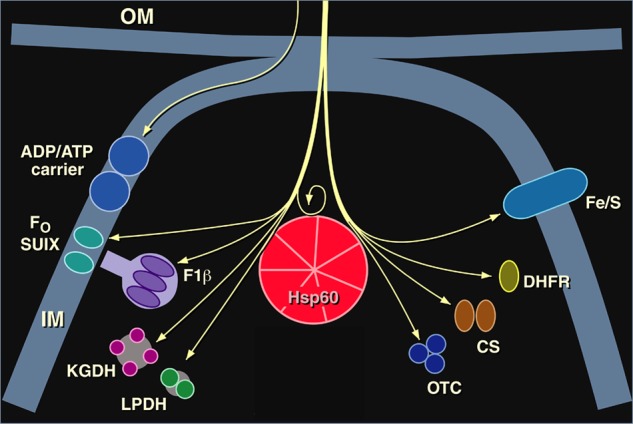
    1. Horwich A. L., Fenton W. A., Williams K. R., Kalousek F., Kraus J. P., Doolittle R. F., Konigsberg W., Rosenberg L. E. (1984) Structure and expression of a cDNA for the nuclear coded precursor of human mitochondrial ornithine transcarbamylase. Science 224, 1068–1074 - PubMed
    1. Horwich A. L., Kalousek F., Mellman I., Rosenberg L. E. (1985) A leader peptide is sufficient to direct mitochondrial import of a chimeric protein. EMBO J. 4, 1129–1135 - PMC - PubMed
    1. Cheng M. Y., Pollock R. A., Hendrick J. P., Horwich A. L. (1987) Import and processing of human ornithine transcarbamoylase precursor by mitochondria from Saccharomyces cerevisiae. Proc. Natl. Acad. Sci. U.S.A. 84, 4063–4067 - PMC - PubMed
    1. Eilers M., Schatz G. (1986) Binding of a specific ligand inhibits import of a purified precursor protein into mitochondria. Nature 322, 228–232 - PubMed

Personal name as subject

LinkOut - more resources