. 2013 Aug 16;288(33):23704-15.
doi: 10.1074/jbc.M113.481572. Epub 2013 Jun 26.

Membrane damage by an α-helical pore-forming protein, Equinatoxin II, proceeds through a succession of ordered steps

Affiliations

Membrane damage by an α-helical pore-forming protein, Equinatoxin II, proceeds through a succession of ordered steps

Nejc Rojko et al. J Biol Chem. .

Abstract

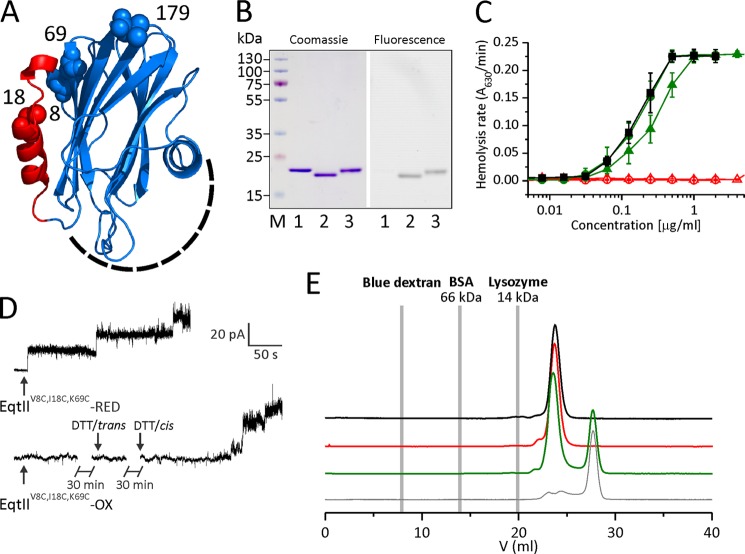
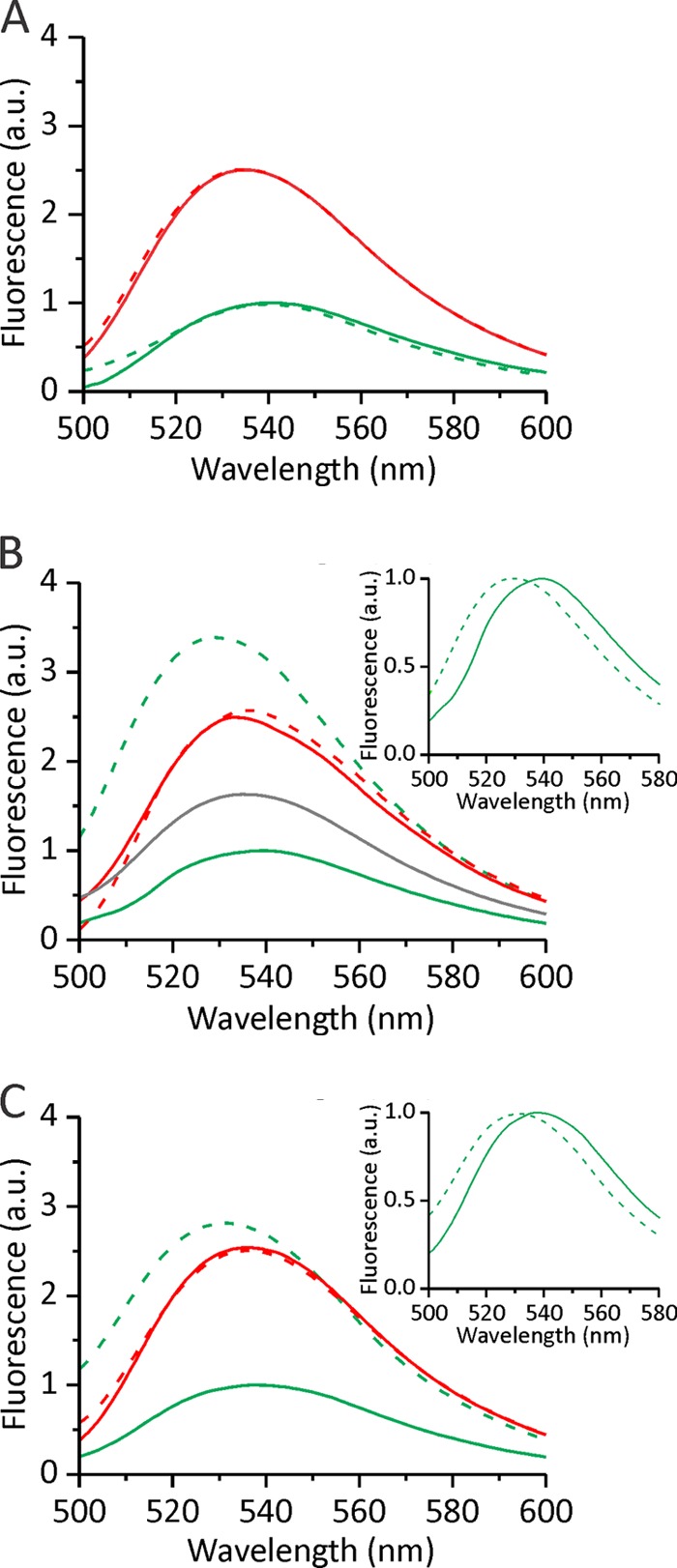
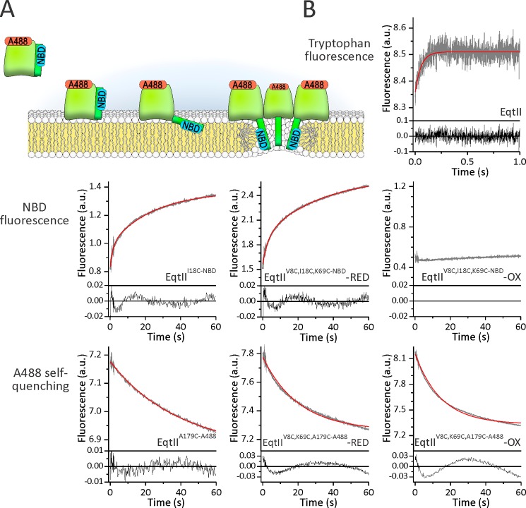
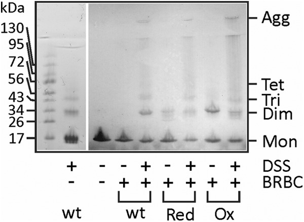
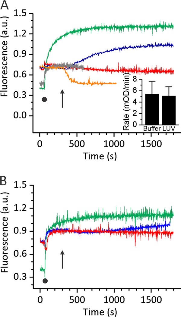
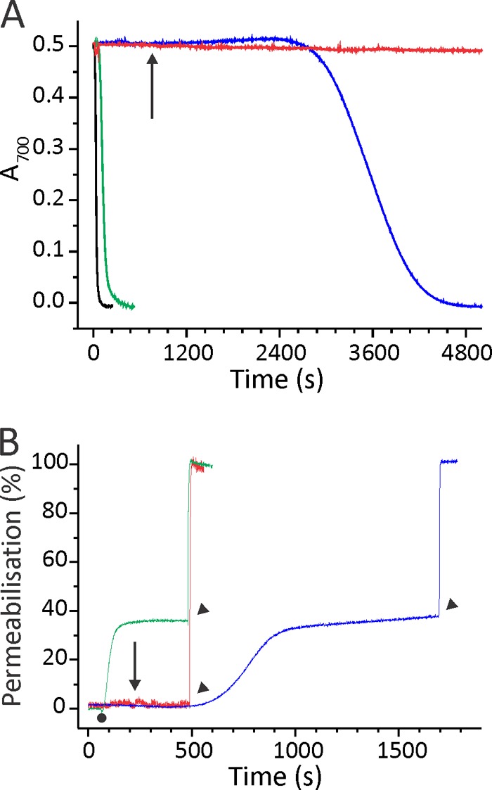
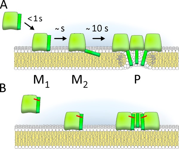
Actinoporin equinatoxin II (EqtII) is an archetypal example of α-helical pore-forming toxins that porate cellular membranes by the use of α-helices. Previous studies proposed several steps in the pore formation: binding of monomeric protein onto the membrane, followed by oligomerization and insertion of the N-terminal α-helix into the lipid bilayer. We studied these separate steps with an EqtII triple cysteine mutant. The mutant was engineered to monitor the insertion of the N terminus into the lipid bilayer by labeling Cys-18 with a fluorescence probe and at the same time to control the flexibility of the N-terminal region by the disulfide bond formed between cysteines introduced at positions 8 and 69. The insertion of the N terminus into the membrane proceeded shortly after the toxin binding and was followed by oligomerization. The oxidized, non-lytic, form of the mutant was still able to bind to membranes and oligomerize at the same level as the wild-type or the reduced form. However, the kinetics of the N-terminal helix insertion, the release of calcein from erythrocyte ghosts, and hemolysis of erythrocytes was much slower when membrane-bound oxidized mutant was reduced by the addition of the reductant. Results show that the N-terminal region needs to be inserted in the lipid membrane before the oligomerization into the final pore and imply that there is no need for a stable prepore formation. This is different from β-pore-forming toxins that often form β-barrel pores via a stable prepore complex.

Keywords: Actinoporin; Equinatoxin; Erythrocyte; Fluorescence; Kinetics; Membrane; Pore Forming; Toxins.


Figures

FIGURE 1.
FIGURE 1.
Properties of engineered EqtII mutant. A, three-dimensional model of EqtII. The N-terminal region, which is α-helical in the lipid membrane environment (19, 24), is shown in red. Residues at positions 8, 18, 69, and 179 are shown with spheres and denoted by the residue number. The membrane binding region is located on the bottom of the molecule and is composed of amino acids from the C-terminal helix and large loops at the bottom of the molecule (schematically denoted by a dashed line). B, SDS-PAGE of EqtIIV8C,I18C,K69C-NBD (left) and the same gel under UV light before Coomassie Blue staining (right). 1, wild type EqtII; 2, EqtIIV8C,I18C,K69C-NBD-OX; 3, EqtIIV8C,I18C,K69C-NBD-RED; M, molecular weight marker. EqtIIV8C,I18C,K69C-NBD-OX shows a gel mobility shift due to the disulfide bridge formed between Cys-8 and Cys-69. C, the rate of hemolysis of the wild type (black squares), EqtIIV8C,I18C,K69C (triangles), and EqtIIV8C,I18C,K69C-NBD (circles) was measured turbidimetrically at 630 nm by using a microplate reader. Shown are reduced (solid symbols and green) and oxidized (open symbols and red) variants of EqtIIV8C,I18C,K69C. n = 10–18, average ± S.D. (error bars). Oxidized mutants are unable to transfer the N-terminal helix to the lipid environment, thus preventing their hemolytic activity. D, activity of the EqtIIV8C,I18C,K69C in PLB. PLB were formed of 1,2-diphytanoyl-sn-glycerophosphocholine and 20% (w/w) SM. The buffer was 10 mm Tris-HCl, 100 mm KCl, pH 8.0, on both sides of the membrane. EqtIIV8C,I18C,K69C-RED or EqtIIV8C,I18C,K69C-OX was added at a 2 nm (RED) and 10 nm (OX) final concentration to the cis side, where a constant voltage +40 mV was applied. 1 mm DTT was added into the trans chamber 30 min after the addition of EqtIIV8C,I18C,K69C-OX and to the cis side after another 30-min incubation (denoted by arrows). Representative traces of at least two independent experiments are shown. E, gel filtration chromatography of the wild type (black), EqtIIV8C,I18C,K69C-NBD-OX (red), and EqtIIV8C,I18C,K69C-NBD-RED (green) on a Superdex HR200 column. EqtII mutants eluted as a single peak, showing no sign of aggregation in solution. The buffer used was 20 mm phosphate buffer, 300 mm NaCl, pH 7.2, at 0.5 ml/min. The elution volume of markers is denoted by gray lines. EqtIIV8C,I18C,K69C-NBD-RED was preincubated with DTT, which resulted in the additional peak in the chromatogram. 30 mm DTT injection in the running buffer is shown as a control by the thin gray trace.
FIGURE 2.
FIGURE 2.
NBD steady-state fluorescence emission spectra. NBD fluorescence of ∼250 nm EqtIIV8C,I18C,K69C-NBD in 140 mm NaCl, 20 mm Tris-HCl, 1 mm EDTA, pH 8.5, was measured in solution (solid line) and in the presence of liposomes of different composition at an L/T of 100 (dashed line). The excitation wavelength was set to 470 nm. The spectra were acquired under constant stirring and at 25 °C. The spectra presented are representative of 2–3 experiments. The insets in B and C show normalized spectra of reduced mutant in the absence (solid line) and presence (dashed line) of liposomes and ghosts, respectively. Green, EqtIIV8C,I18C,K69C-NBD-RED; red, EqtIIV8C,I18C,K69C-NBD-OX. A, DOPC LUV; B, DOPC/SM (1:1) LUV and 5-NO-PC/DOPC/SM (2:3:5) (gray); C, BRBC ghosts. a.u., arbitrary units.
FIGURE 3.
FIGURE 3.
Comparison of binding, insertion of the N-terminal region, and oligomerization by stopped-flow fluorescence measurements. A, a pore-forming mechanism of EqtII and different steps that were studied. Approximate positions of residues at positions 18 and 179 are presented by blue and red labels, respectively. B, fluorescence traces of different probes as denoted. The proteins that were used in each experiment are noted. The concentration of lipids was 10 μm. DOPC/SM (1:1) LUV in 140 mm NaCl, 20 mm Tris-HCl, 1 mm EDTA, pH 8.5, were used. Protein concentration was 100 nm to achieve an L/T ratio of 100. A minimum of four single injections were averaged to obtain a single fluorescent trace (gray). Residuals of the measured data to an exponential fit are shown below the traces. The red trace shows fit to a mono- or biexponential equation (as reported under “Results”).
FIGURE 4.
FIGURE 4.
Oligomerization at the surface of the erythrocytes. 0.25 nmol of protein was incubated with BRBC suspension. After a 10-min incubation and the addition of DSS, BRBC with bound cross-linked toxin were pelleted. Electrophoresis buffer was added to the pelleted erythrocytes, and proteins were resolved by SDS-PAGE and blotted with rabbit anti-EqtII serum. Mon, monomer; Dim, dimer; Tri, trimer; Tet, tetramer; Agg, high molecular weight aggregates; wt, wild-type EqtII; Red, EqtIIV8C,I18C,K69C-RED; Ox, EqtIIV8C,I18C,K69C-OX. The left panel shows EqtII cross-linked in solution in the absence of BRBC. The gel was stained with Coomassie Blue.
FIGURE 5.
FIGURE 5.
Kinetic measurements of N-terminal helix insertion into the lipid environment. NBD fluorescence of 250 nm EqtIIV8C,I18C,K69C-NBD in reduced and oxidized form was measured at 538 nm. After 60 s, liposomes or ghosts (denoted by a black dot) were added at an L/T ratio of 100. Green, EqtIIV8C,I18C,K69C-NBD-RED in the presence of DOPC/SM LUV or ghosts; red, EqtIIV8C,I18C,K69C-NBD-OX in the presence of DOPC/SM LUV or ghosts; blue, EqtIIV8C,I18C,K69C-NBD-OX in the presence of DOPC/SM LUV or ghosts and reduced with final 2 mm DTT at 300 s (denoted by an arrow); orange, EqtIIV8C,I18C,K69C-NBD-OX in solution and reduced with final 2 mm DTT at 300 s (denoted by an arrow). The N-terminal helix insertion shows slower kinetics when the disulfide bridge is reduced after the protein fully binds to the lipid membrane. A, LUV composed of DOPC/SM; B, BRBC ghosts. The traces are representative of three independent experiments. Inset in A, the rate of the disulfide bond cleavage as monitored by the NTSB assay for EqtIIV8C,I18C,K69C in solution and when bound to LUV. Error bars, S.D. a.u., arbitrary units.
FIGURE 6.
FIGURE 6.
Permeabilizing activity of EqtIIV8C,I18C,K69C-NBD. A, the restoration of hemolytic activity of EqtIIV8C,I18C,K69C-NBD-OX by the DTT addition. BRBC were stirred in hemolysis buffer at 25 °C. Proteins were added to a final concentration of 7.5 nm. The figure is representative of at least six independent experiments. The reductant DTT was added at a final 2 mm concentration to EqtIIV8C,I18C,K69C-NBD-OX already bound to the membrane (denoted by an arrow). B, permeabilizing activity on calcein-loaded ghosts. Ghosts were stirred in vesicle buffer at 25 °C. The excitation and emission wavelengths were set to 485 and 520 nm, respectively. The addition of 50 nm EqtIIV8C,I18C,K69C-NBD-RED or EqtIIV8C,I18C,K69C-NBD-OX is denoted by a small dot. The percentage of permeabilization was determined at the end of the assay by comparing the fluorescence with the maximal one, obtained by the addition of 2 mm final Triton X-100 (denoted by arrowheads). EqtIIV8C,I18C,K69C-NBD-OX was also reduced after binding to ghosts with final 2 mm DTT (denoted by an arrow) to show that permeabilizing activity is slower in comparison with the protein reduced before binding to ghosts. The figures are representative of four independent experiments. The color code is the same for both panels: green, EqtIIV8C,I18C,K69C-NBD-RED; red, EqtIIV8C,I18C,K69C-NBD-OX; blue, EqtIIV8C,I18C,K69C-NBD-OX reduced with final 2 mm DTT after the protein was bound to the membranes; black, the wild type EqtII.
FIGURE 7.
FIGURE 7.
Pore-forming mechanism of EqtII. A, pore formation proceeds through membrane binding (M1 state (25)), N-terminal helix insertion in the membrane (M2), and oligomerization to a final pore (P). B, mutant in which cysteines at positions 8 and 69 locked N-terminal to the body of the molecule could bind to the membrane and oligomerize but could not form the pore.

Similar articles

Cited by

References

    1. Parker M. W., Feil S. C. (2005) Pore-forming protein toxins. From structure to function. Prog. Biophys. Mol. Biol. 88, 91–142 - PubMed
    1. Anderluh G., Lakey J. H. (2008) Disparate proteins use similar architectures to damage membranes. Trends Biochem. Sci. 33, 482–490 - PubMed
    1. Dalla Serra M., Tejuca Martinez M. (2011) Pore-forming toxins. eLS, DOI: 10.1002/9780470015902.a0002655.pub2 - DOI
    1. Heuck A. P., Tweten R. K., Johnson A. E. (2001) β-barrel pore-forming toxins. Intriguing dimorphic proteins. Biochemistry 40, 9065–9073 - PubMed
    1. Mueller M., Grauschopf U., Maier T., Glockshuber R., Ban N. (2009) The structure of a cytolytic α-helical toxin pore reveals its assembly mechanism. Nature 459, 726–730 - PubMed

Publication types

MeSH terms

LinkOut - more resources